Skip directly to site content Skip directly to page options Skip directly to A-Z link Skip directly to A-Z link Skip directly to A-Z link
Volume 14, Number 8—August 2008
Dispatch

Pathogenicity of Highly Pathogenic Avian Influenza Virus (H5N1) in Adult Mute Swans

Donata Kalthoff*, Angele Breithaupt*, Jens P. Teifke*, Anja Globig*, Timm Harder*, Thomas C. Mettenleiter*, and Martin Beer*Comments to Author 
Author affiliations: *Friedrich-Loeffler-Institut, Greifswald-Insel Riems, Germany;

Main Article

Figure 2

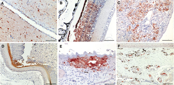
Figure 2 - Immunohistochemical analysis for nucleoprotein of avian influenza virus. Tissue sections were stained by using the avidin-biotin-peroxidase complex method, 3-amino-9-ethylcarbazole (red), and hematoxylin (blue). A) Brain, cerebrum: numerous glial cells, neurons and ependymal cells stain positive for influenza virus antigen (scale bar = 200 μm). B) Eye, retina: cells of the pigmented epithelial layer, photoreceptor cells, and cells of the outer and inner nuclear layers are po

Figure 2. Immunohistochemical analysis for nucleoprotein of avian influenza virus. Tissue sections were stained by using the avidin-biotin-peroxidase complex method, 3-amino-9-ethylcarbazole (red), and hematoxylin (blue). A) Brain, cerebrum: numerous glial cells, neurons and ependymal cells stain positive for influenza virus antigen (scale bar = 200 μm). B) Eye, retina: cells of the pigmented epithelial layer, photoreceptor cells, and cells of the outer and inner nuclear layers are positive for the nucleoprotein of influenza virus (scale bar = 100 μm). C) Liver: subadjacent to the capsule there is hepatocyte degeneration and necrosis around a congested central vein (scale bar = 100 μm). D) Skin: keratinized layer of the feather follicular epithelium shows focal necrosis with intense nuclear and cytoplasmic immunostaining (scale bar =100 μm). E) Nasal cavity: focal intraepithelial necrosis of the mucocutaneous membrane associated with influenza virus infection (scale bar = 50 μm). F) Nasal concha: numerous submucosal arterioles and venules display strong endothelial staining, which partially extends into the media of the vessels (scale bar = 100 μm).

Main Article

Page created: July 12, 2010
Page updated: July 12, 2010
Page reviewed: July 12, 2010
The conclusions, findings, and opinions expressed by authors contributing to this journal do not necessarily reflect the official position of the U.S. Department of Health and Human Services, the Public Health Service, the Centers for Disease Control and Prevention, or the authors' affiliated institutions. Use of trade names is for identification only and does not imply endorsement by any of the groups named above.
file_external