Volume 15, Number 11—November 2009
Letter
Rickettsia africae Infection in Man after Travel to Ethiopia
Cite This Article
Citation for Media
To the Editor: The first human case of African tick-bite fever was described in 1992 as occurring in Zimbabwe. The causative agent was identified as a new serotype of the spotted fever group (SFG) rickettsiae and named Rickettsia africae (1). These findings confirmed observations made by Pijper in the 1930s, which suggested that there were 2 different kinds of human SFG rickettsioses in sub-Saharan Africa: Mediterranean spotted fever caused by R. conorii and transmitted by Rhipicephalus species, ticks of dogs, and African tick-bite fever caused by R. africae and transmitted by Amblyomma species, ticks of cattle and wild ungulates. African tick-bite fever has subsequently been diagnosed in patients from several other sub-Saharan countries and also from the West Indies (2,3).
In a recent analysis of the spectrum of diseases among returning travelers, tick-borne spotted fever was (after malaria) the second most frequent cause of systemic febrile illness among those returning from sub-Saharan Africa. It occurred more frequently than typhoid fever and dengue fever (4). The following case description reports an infection with R. africae in a man in France who recently returned from Ethiopia.
Figure
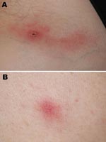
Figure. Inoculation eschar on left inguinal fold (A) and vesicular skin lesion (B) in a traveler recently returned to France from Ethiopia.
On November 4, 2005, a 62-year-old French man sought care at the Medical Center of the Institut Pasteur in Paris for fever, along with chills, headache, neck and shoulder pain, and fatigue over the previous 4 days. At the onset of these symptoms he had noticed dark nodular lesions on his neck and his left groin followed 2 days later by a slightly painful eruption on his arms and his trunk. He had spent a month in southwest Ethiopia, north of Kelem near the Sudanese border, and returned to France on October 26, 2005. While in Ethiopia, he had assisted with a production of a documentary film about an Ethiopian tribe and had been in contact with cattle in the villages. He had not noticed any tick bites. On physical examination he had a fever of 38°C, a nodular lesion with a central dark crust on his neck, a second lesion on his left inguinal fold (Figure, panel A), and a vesicular eruption on his arms and his trunk (Figure, panel B). Leukocyte count was 3,200, including 1,869 neutrophils and 867 lymphocytes. The platelet level was 174,000/mm3. The C-reactive protein level was 28.3 mg/L. The aspartate aminotransferase level was slightly elevated. The patient was treated with doxycycline 200 mg/day for 1 week for suspected African tick-bite fever. Follow-up showed a quick recovery from his symptoms except for fatigue that persisted for ≈1 month.
A commercial immunofluorescence assay for R. conorii and R. typhi immunoglobulin G performed both on an initial blood sample and a second sample taken 1 week later were negative. A blood sample and a biopsy specimen of the inguinal eschar were sent to the National Reference Center of Rickettsiae in Marseille, France. Although cellular culture of both specimens and molecular testing of the blood sample were negative, PCR for the sequences of citrate synthase (GenBank accession no. RAU59733, 93.1% homology) and rickettsial OmpA (GenBank accession no. RAU83436, 99.3% homology) applied on the skin biopsy detected R. africae and confirmed the diagnosis of African tick-bite fever.
From 1969 to 1971, SFG rickettsiae were isolated from Amblyomma spp. ticks collected in Ethiopia. They were regarded as R. conorii or as closely related bacteria (5). Later, more specific tests using western immunoblots with monoclonal antibodies showed that these rickettsiae differed from R. conorii (6). In 1992 SFG rickettsiae isolated from Amblyomma ticks collected in Zimbabwe and from the blood of a patient in Zimbabwe were compared to R. conorii, to other pathogenic SFG rickettsiae, and to a SFG rickettsia isolated from an Amblyomma spp. tick in Ethiopia 20 years before. The SFG rickettsia isolates from Ethiopia were identical to isolates obtained in Zimbabwe from the Amblyomma ticks and the patient’s blood and were different from R. conorii and other pathogenic SFG rickettsiae. This new serotype of SFG rickettsiae was named R. africae (1,7). A recent study confirmed the presence of R. africae in ticks collected in Ethiopia, as well as R. aeschlimanii (8). Thus, evidence of R. africae in Ethiopia has been known for a long time.
The geographic distribution of African tick-bite fever is related to the presence of Amblyomma spp. ticks, vectors and reservoirs of R. africae. Consequently African tick-bite fever should also be considered as a possible diagnosis in patients with febrile illness returning from countries where R. africae has been detected in Amblyomma ticks, even if a human infection has not yet been reported (9,10).
References
- Kelly PJ, Beati L, Matthewman LA, Mason PR, Dasch GA, Raoult D. A new pathogenic spotted fever group rickettsia from Africa. J Trop Med Hyg. 1994;97:129–37.PubMedGoogle Scholar
- Raoult D, Fournier PE, Fenollar F, Jensenius M, Prioe T, de Pina JJ, Rickettsia africae, a tick-borne pathogen in travelers to sub-Saharan Africa. N Engl J Med. 2001;344:1504–10. DOIPubMedGoogle Scholar
- Ndip LM, Bouyer DH, Travassos Da Rosa AP, Titanji VP, Tesh RB, Walker DH. Acute spotted fever rickettsiosis among febrile patients, Cameroon. Emerg Infect Dis. 2004;10:432–7.PubMedGoogle Scholar
- Freedman DO, Weld LH, Kozarsky PE, Fisk T, Robins R, von Sonnenburg F, Spectrum of disease and relation to place of exposure among ill returned travelers. N Engl J Med. 2006;354:119–30. DOIPubMedGoogle Scholar
- Burgdorfer W, Ormsbee RA, Schmidt ML, Hoogstraal H. A search for the epidemic typhus agent in Ethiopian ticks. Bull World Health Organ. 1973;48:563–9.PubMedGoogle Scholar
- Walker DH, Liu QH, Yu XJ, Li H, Taylor C, Fenq HM. Antigenic diversity of Rickettsia conorii. Am J Trop Med Hyg. 1992;47:78–86.PubMedGoogle Scholar
- Kelly PJ, Matthewman LA, Beati L, Raoult D, Mason P, Dreary M, African tick bite fever: a new spotted fever group under an old name. Lancet. 1992;340:982–3.Medline DOIGoogle Scholar
- Mura A, Socolovschi C, Ginesta J, Lafrance B, Magnan S, Rolain JM, Molecular detection of spotted fever group rickettsiae in ticks from Ethiopia and Chad. Trans R Soc Trop Med Hyg. 2008;102:945–9. DOIPubMedGoogle Scholar
- Parola P, Raoult D. Ticks and tickborne bacterial diseases in humans: an emerging infectious threat. Clin Infect Dis. 2001;32:897–928. DOIPubMedGoogle Scholar
- Parola P, Barre N. Rickettsia africae, agent de la fièvre à tique africaine: un pathogène émergent dans les Antilles et l’île de la Réunion. Bull Soc Pathol Exot. 2004;97:193–8.PubMedGoogle Scholar
Figure
Cite This ArticleRelated Links
Table of Contents – Volume 15, Number 11—November 2009
| EID Search Options |
|---|
|
|
|
|
|
|
Please use the form below to submit correspondence to the authors or contact them at the following address:
Paul H. Consigny, Centre Médical, Institut Pasteur, 28, rue du Docteur Roux, 75724 Paris Cedex 15, France
Top