Volume 15, Number 3—March 2009
Dispatch
Human Bocavirus and KI/WU Polyomaviruses in Pediatric Intensive Care Patients
Cite This Article
Citation for Media
Abstract
We evaluated the prevalence of human bocavirus and KI and WU polyomaviruses in pediatric intensive care patients with and without lower respiratory tract infection (LRTI). The prevalence of these viruses was 5.1%, 0%, and 2.6%, respectively, in children with LRTI and 4.8%, 4.8%, and 2.4%, respectively, in those without LRTI.
Through use of molecular diagnostic tests such as real-time PCR in the clinical setting, our scope of etiologic viral agents of lower respiratory tract infection (LRTI) has increased. Respiratory viruses can now be detected in most pediatric intensive care patients with LRTI (1). Recently, 3 new viruses were described: human bocavirus (HBoV) and KI (KIPyV) and WU (WUPyV) polyomaviruses (2–4). These viruses were first identified in respiratory samples obtained from children with respiratory tract infections. An association between the viruses and respiratory tract symptoms was postulated, but, to date, evidence supporting that association is incomplete (5–7). This study evaluates the prevalences of HBoV, KIPyV, and WUPyV infections in pediatric intensive care patients with acute respiratory insufficiency caused by LRTI.
Patients <5 years of age who were admitted for LRTI to the pediatric intensive care unit (PICU) of Wilhelmina Children’s Hospital, Utrecht, the Netherlands, were enrolled from October through May during 2005–2008. Patients were excluded if they had any of the following: asthma exacerbation, immunocompromised state, indication for antimicrobial drugs other than for LRTI, and repeated PICU admission for LRTI during the study period. Control group participants were children <18 years of age (median 2.2 years) who were admitted to the PICU from October 2005 through March 2006 for reasons other than LRTI.
Clinical data were obtained by using standardized forms to extract data from electronic charts. Underlying illnesses were defined as chronic pulmonary disease, congenital heart disease, immunodeficiency, malignancy, neurologic disease, or gastrointestinal disease (8). To assess the severity of illness, we used the lowest ratio during the first 24 hours of the partial pressure of oxygen in arterial blood (PaO2) to the inspired oxygen fraction (FiO2). These ratios were acquired from the Pediatric Intensive Care Evaluation database, which contains validated clinical data for all Dutch PICU admissions.
Nasopharyngeal aspirates were collected from all patients in the LRTI group as part of the investigation of their illnesses. In the control group, nasopharyngeal aspirates were taken from intubated patients, and throat swabs were taken from extubated children as part of routine surveillance to identify transmission of respiratory syncytial virus (RSV). Because RSV surveillance was conducted as part of normal patient care, patient consent/ethical approval was not needed, according to the Medical Ethical Research Council of our institution.
Specimens from patients in the LRTI group were initially examined for RSV, influenza viruses, parainfluenza viruses, adenoviruses, rhinoviruses, coronaviruses, human metapneumovirus, and Mycoplasma pneumoniae by using real-time PCR as previously described (1,9,10). Specimens from patients in the control group were initially examined for RSV. All samples were retrospectively tested for HBoV, KIPyV, and WUPyV also by using real-time PCR as previously described (11,12). After nucleic acid extraction using the MagNA Pure LC 1.0 nucleic acid isolation system (Roche Diagnostics, Rotkreuz, Switzerland), amplification was carried out in a 25-μL reaction mixture on a 7500 Fast Real-Time PCR System (Applied Biosystems, Foster City, CA, USA). Positive controls for the KIPyV and WUPyV PCR were provided by S. Bialasiewicz and T.P. Sloots, University of Queensland, Queensland, Australia, and the positive control for HBoV was provided by T. Allander, Karolinska Institute, Stockholm, Sweden. Internal control viruses were used to monitor efficient extraction and amplification. Real-time PCR results were expressed in cycle threshold (Ct) values. Ct values are inversely correlated with viral load; i.e., low Ct values indicate high viral loads.
Of 90 LRTI patients enrolled, 78 (86.7%) had sufficient material stored for HBoV, KIPyV, and WUPyV testing. Eighty-eight control patients were enrolled, of which 83 (94.3%) had sufficient material stored to be included. Table 1 provides patients’ demographic and clinical characteristics. The main clinical conditions of control patients were cardiac disease requiring surgery (33.7%), trauma (8.4%), sepsis (8.4%), and upper respiratory tract infection (8.4%). A total of 57 (68.7%) nasopharyngeal aspirates and 26 (31.3%) throat swabs from the 83 patients who had sufficient samples were tested.
In LRTI patients, HBoV was found in 4 (5.1%) and WUPyV in 2 (2.6%) of the 78 patients. No samples tested positive for KIPyV. Table 2 shows Ct values and clinical characteristics for LRTI patients whose samples were positive as well as for controls whose samples were positive for these viruses. Other respiratory viruses were found in 70 (89.7%) of the 78 children. RSV was found in 52 (66.7%), influenza viruses in 3 (3.8%), parainfluenza viruses in 2 (2.6%), adenoviruses in 4 (5.1%), rhinoviruses in 20 (25.6%), coronaviruses in 6 (7.7%), human metapneumovirus in 5 (6.4%), and Mycoplasma pneumoniae in 1 (1.3%) of the patients. Multiple respiratory viruses were found in 3 of the 4 LRTI patients with HBoV infection and in both patients with WuPyV infection (Table 2). One patient had a single infection with HBoV (i.e., no other virus was detected). This patient was born at 31 weeks of gestational age and had a history of a grade IV idiopathic respiratory distress syndrome. She was admitted to the PICU at 19 months of age with a severe LRTI. Bacterial throat and blood cultures remained negative.
In the control group, HBoV was found in samples from 4 (4.8%) patients, KIPyV was found in 4 (4.8%), and WUPyV was present in 2 (2.4%) samples from the 83 patients whose samples could be tested. One patient was found to have a co-infection with HBoV and KIPyV.
Figure
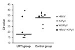
Figure. Cycle threshold (Ct) values of lower respiratory tract infection (LRTI) and control patients with human bocavirus (HBoV), KI polyomavirus (KIPyV), and WU polyomavirus (WUPyV) infections. LRTI patients are those admitted to...
Median Ct values for HBoV, KIPyV, and WUPyV combined were 18.1 (interquartile range [IQR] 20.4) for the LRTI group and 34.4 (IQR 5.1) for the control group (p = 0.09; Figure). The Ct values indicate that, on average, the viral load in the LRTI group might be higher than in the control group.
In the present study, the prevalences of HBoV, KIPyV, and WUPyV in PICU patients with LRTI (n = 78) or without LRTI (n = 83) were similar (5.1%, 0%, 2.6%; and 4.8%, 4.8%, 2.4%, respectively). Most HBoV- and KIPyW-positive LRTI patients were co-infected with other viruses. One LRTI patient with a HBoV single infection was identified. In this patient, HBoV was present in a high quantity.
Two limitations of our study deserve further discussion. First, LRTI patients were younger than controls (LRTI, 100% <5 years; controls, 40% >5 years). However, the positivity rates for HBoV, KIPyV, and WUPyV in control children <5 years were similar to rates of control children >5 years of age (6/49 vs. 3/34, respectively). Hence, the influence of this limitation is likely minor. Studies have also shown that the highest incidence of KIPyV/WUPyV infection occurs in children ≈1 year of age, slightly older than the children in our LRTI group (13–15). The young age of the LRTI group may have resulted in a lower than expected positivity rate for this group. Second, all LRTI patients had nasopharyngeal aspirates taken; however, 68.7% of controls had provided nasopharyngeal aspirates. HBoV and KIPyV/WUPyV infections were more common in controls who had nasopharyngeal aspirate samples taken than in those who had throat swab samples taken (8/57 vs. 1/26). This difference in positivity for nasopharyngeal aspirates strengthens our conclusion that these viruses are not found more frequently in PICU children with LRTI.
Sampling errors make precise quantification of viral loads difficult. Nevertheless, in the LRTI group, low Ct values, which indicate high viral loads, were found in nasopharyngeal samples taken from 3 patients infected with HBoV (Ct ≈ 15) and from 1 patient infected with WUPyV (Ct = 19). Ct values found in nasopharyngeal samples from patients in the control group were much higher, 32–39. A possible explanation for this difference is that high viral loads in the young LRTI population represent symptomatic primary infection, whereas the low viral load in the older controls might represent asymptomatic long-term shedding. Further studies are needed to show the clinical implications of infections with these viruses.
Prevalences of HBoV, KIPyV, and WUPyV infections in children in the PICU is low (≈<5% for LRTI patients and controls), and these agents are unlikely to be a major cause of LRTI at the PICU. However, HBoV might be pathogenic in some PICU patients because 1 person with a HBoV single infection in a high quantity was identified. Further studies using quantitative viral detection are needed to investigate the probability that HBoV, KIPyV, and WUPyV represent etiologic agents of LRTI.
Ms van de Pol is a PhD candidate at the University Medical Center Utrecht, Utrecht, the Netherlands. Her research focuses on the clinical implications of nucleic acid amplification tests for respiratory viruses at the pediatric intensive care unit.
Acknowledgments
We thank Marco Viveen, Ramona Zenhorst, and Ilse Overdevest for technical assistance; Niels Lastdrager for reviewing electronic charts; and Idse Visser for providing PaO2/FiO2 ratios.
This work was supported by an “Alexandre Suerman program” MD/PhD grant from the University Medical Center Utrecht, Utrecht, the Netherlands.
References
- van de Pol AC, Wolfs TF, Jansen NJ, van Loon AM, Rossen JW. Diagnostic value of real-time polymerase chain reaction to detect viruses in young children admitted to the paediatric intensive care unit with lower respiratory tract infection. Crit Care. 2006;10:R61. DOIPubMedGoogle Scholar
- Allander T, Tammi MT, Eriksson M, Bjerkner A, Tiveljung-Lindell A, Andersson B. Cloning of a human parvovirus by molecular screening of respiratory tract samples. Proc Natl Acad Sci U S A. 2005;102:12891–6. DOIPubMedGoogle Scholar
- Allander T, Andreasson K, Gupta S, Bjerkner A, Bogdanovic G, Persson MA, Identification of a third human polyomavirus. J Virol. 2007;81:4130–6. DOIPubMedGoogle Scholar
- Gaynor AM, Nissen MD, Whiley DM, Mackay IM, Lambert SB, Wu G, Identification of a novel polyomavirus from patients with acute respiratory tract infections. PLoS Pathog. 2007;3:e64. DOIPubMedGoogle Scholar
- McIntosh K. Human bocavirus: developing evidence for pathogenicity. J Infect Dis. 2006;194:1197–9. DOIPubMedGoogle Scholar
- Sloots TP, Whiley DM, Lambert SB, Nissen MD. Emerging respiratory agents: new viruses for old diseases? J Clin Virol. 2008;42:233–43. DOIPubMedGoogle Scholar
- van der Zalm MM, Rossen JW, van Ewijk BE, Wilbrink B, van Esch PC, Wolfs TF, Prevalence and pathogenicity of WU and KI polyomaviruses in children, the Netherlands. Emerg Infect Dis. 2008;14:1787–9. DOIPubMedGoogle Scholar
- Arnold JC, Singh KK, Spector SA, Sawyer MH. Human bocavirus: prevalence and clinical spectrum at a children’s hospital. Clin Infect Dis. 2006;43:283–8. DOIPubMedGoogle Scholar
- van de Pol AC, van Loon AM, Wolfs TF, Jansen NJ, Nijhuis M, Breteler EK, Increased detection of respiratory syncytial virus, influenza viruses, parainfluenza viruses, and adenoviruses with real-time PCR in samples from patients with respiratory symptoms. J Clin Microbiol. 2007;45:2260–2. DOIPubMedGoogle Scholar
- van Elden LJ, van Loon AM. van der BA, Hendriksen KA, Hoepelman AI, van Kraaij MG, et al. Applicability of a real-time quantitative PCR assay for diagnosis of respiratory syncytial virus infection in immunocompromised adults. J Clin Microbiol. 2003;41:4378–81. DOIPubMedGoogle Scholar
- Allander T, Jartti T, Gupta S, Niesters HG, Lehtinen P, Osterback R, Human bocavirus and acute wheezing in children. Clin Infect Dis. 2007;44:904–10. DOIPubMedGoogle Scholar
- Bialasiewicz S, Whiley DM, Lambert SB, Gould A, Nissen MD, Sloots TP. Development and evaluation of real-time PCR assays for the detection of the newly identified KI and WU polyomaviruses. J Clin Virol. 2007;40:9–14. DOIPubMedGoogle Scholar
- Abed Y, Wang D, Boivin G. WU polyomavirus in children, Canada. Emerg Infect Dis. 2007;13:1939–41.PubMedGoogle Scholar
- Han TH, Chung JY, Koo JW, Kim SW, Hwang ES. WU polyomavirus in children with acute lower respiratory tract infections, South Korea. Emerg Infect Dis. 2007;13:1766–8.PubMedGoogle Scholar
- Norja P, Ubillos I, Templeton K, Simmonds P. No evidence for an association between infections with WU and KI polyomaviruses and respiratory disease. J Clin Virol. 2007;40:307–11. DOIPubMedGoogle Scholar
Figure
Tables
Cite This ArticleTable of Contents – Volume 15, Number 3—March 2009
| EID Search Options |
|---|
|
|
|
|
|
|
Please use the form below to submit correspondence to the authors or contact them at the following address:
Alma C. van de Pol, Wilhelmina Children’s Hospital, University Medical Center Utrecht, Lundlaan 6, 3584 EA Utrecht, the Netherlands;
Top