Volume 15, Number 7—July 2009
Letter
Chitinophaga terrae Bacteremia in Human
Cite This Article
Citation for Media
To the Editor: The genus Chitinophaga, first described by Sangkhobol and Skerman in 1981, belongs to the phylum Bacteroidetes (formerly the Cytophaga-Flexibacter-Bacteroides group), which includes filamentous, chitinolytic, gliding bacteria that transform into spherical bodies upon aging (1). This genus contains 10 environmental species that demonstrate similarities in 16S rDNA sequence and in phenotypic and chemotaxonomic data (menaquinone, fatty acids, hydroxy fatty acid, and polyamine) (2–5). Chitinophaga terrae, originally isolated from soil in South Korea, was first described in 2007 (3,4). Here we report a case of bacteremia due to C. terrae in a severely immunosuppressed woman.
On July 31, 2008, a 51-year-old woman was admitted to the emergency department at Nantes University Hospital in Nantes, France, because of a slowly growing left cheek mass associated with weight loss and change of general state. Physical examination showed several cutaneous infiltrated nodules, bilateral axillary adenopathies, and hepatosplenomegaly. Deteriorating renal function led to intermittent hemodialysis. Histopathology of skin and renal biopsies revealed a diffuse, high–grade, large B-cell lymphoma with cutaneous localization. Systemic CHOP (cyclophosphamide, doxorubicin, vincristine, prednisone) chemotherapy and methotrexate intrathecal chemotherapy were begun August 9. She developed bone marrow aplasia 3 days later, shortly followed by the onset of pyrexia. Corynebacterium pseudotuberculosis was isolated from 3 blood samples, 2 drawn using the central venous catheter (CVC) and 1 from peripheral blood. Consecutively, serotype O1 Pseudomonas aeruginosa strain was isolated from cultures of urine and fecal specimens. The empirical antimicrobial drug treatment started with piperacillin-tazobactam, ciprofloxacin, and teicoplanin on August 16 and was replaced by imipenem, ciprofloxacin, and teicoplanin on August 21. On August 26, the patient was admitted to the medical intensive care unit (MICU) after indications of toxic epidermal necrolysis. Blood analysis showed pancytopenia with thrombocytopenia (26 × 109/L), anemia (hemoglobin 8.7 g/dL), and profound leukopenia (0.01 × 10⁹/L). Antimicrobial drug treatment was changed to an association of noncytotoxic drugs, i.e., aztreonam, amikacin, and teicoplanin. Despite this broad-spectrum antimicrobial therapy, 4 aerobic blood cultures (1 drawn August 29, 2 on September 2, and 1 on September 3), 2 drawn using the CVC and 2 from a peripheral site, yielded gram-negative bacilli (our laboratory reference no. NTS8639) after 2 days’ incubation. The CVC was removed September 3 and sent to the laboratory for culture. The catheter tip was immersed in 2 mL of brain heart infusion agar, and semiquantitative culture was performed on the blood agar plate using 100 µL of the solution. The culture remained negative.
On September 2, treatment was changed to imipenem, trimethoprim-sulfamethoxazole, and teicoplanin. Additionally, diagnosis of invasive pulmonary aspergillosis led to changing caspofungin prophylaxis to voriconazole. Trimethoprim-sulfamethoxazole treatment was stopped September 9, and imipenem was stopped September 25, a week after bone marrow recovery. The patient was discharged from MICU on October 7.
Figure
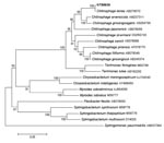
Figure. Neighbor-joining (NJ) tree showing the phylogenetic placement of strain NTS8639 (in boldface) among members of the Chitinophaga terrae species. Twenty-one 16S rRNA gene sequences selected from the GenBank database were aligned...
Yellow-pigmented colonies grew on bromocresol purple lactose agar plate after 48 hours of incubation at 37°C and appeared as thin gram-negative bacilli after gram-staining was performed. The nonfermenting, nonmotile, oxidase-positive bacterium could grow at various pH values (pH 6.0, 7.3, and 8.0) and at different temperatures (30, 37, and 40°C). The semi-automatic Api 20NE gallery (bioMérieux, Marcy l’Etoile, France) identified the strain as Sphingomonas paucimobilis, whereas the ID-GNB card of the VITEK 2 system (bioMérieux) identified the bacterium as Sphingobacterium thalpophilum. The 16S rDNA amplification and sequencing were performed with universal primers 27f and 1378r as previously described (6). The 1366-bp sequence matched that of C. terrae with 100% similarity, according to BIBI (Bioinformatic Bacteria Identification, http://umr5558-sud-str1.univ-lyon1.fr/lebibi/lebibi.cgi) or BLAST (www.ncbi.nlm.nih.gov) analysis. Phylogenetic analysis with either the neighbor-joining or maximum-parsimony algorithm embedded the NTS8639 strain to the genus Chitinophaga and the species C. terrae (Figure). The biochemical characteristics of the bacterium corresponded to those previously described for C. terrae by Kim and Jung (3). The strain reduced nitrate to nitrite, produced N-acetyl-β-glucosamidase, phosphatase, α- and β-galactosidases, α- and β-glucosidases, and assimilated
Disk diffusion tests showed that the bacterium was multiresistant to antimicrobial drugs, including most of the β-lactams, aminoglycosides, fluoroquinolones, colistin, fosfomycin, and tigecyclin. It remained susceptible to amoxicillin-clavulanate, ticarcillin-clavulanate, carbapenems, and trimethoprim-sulfamethoxazole. Interpretation of the susceptibility test was impossible before 48 hours incubation, i.e., 4 days after the first positive blood culture.
We describe in this report a case of human bacteremia due to C. terrae. This environmental organism behaving as an opportunistic pathogen was able to produce infection in a severely immunosuppressed woman. The source of bacteremia was not clearly established. Catheter-related bacteremia was not confirmed by culture of the CVC tip sent to the laboratory on September 3. Virulence factors contributing to the pathogenicity of C. terrae have not yet been well defined. The infection could have been favored by the immunosuppressive therapy, the profound leukopenia, and the extensive cutaneous detachment subsequently associated with methotrexate overdose in this patient. Unlike most susceptible environmental organisms, this bacterium probably was assisted by its multiresistance to antimicrobial drugs in producing infection. The intrinsic or acquired resistance of C. terrae to antimicrobial drugs has not yet been fully elucidated. The lack of commercially available biochemical gallery databases makes correct identification of this environmental organism difficult. It also underlines the usefulness of 16S rDNA sequencing for identification of unusual gram-negative bacilli isolated from immunocompromised hosts (7).
References
- Shangkhobol V, Skerman VBD. Chitinophaga, a new genus of chitinolytic myxobacteria. Int J Syst Evol Microbiol. 1981;31:285–93. DOIGoogle Scholar
- Kämpfer P, Young CC, Sridhar KR, Arun AB, Lai WA, Shen FT, Transfer of [Flexibacter] sancti, [Flexibacter] filiformis, [Flexibacter] japonensis and [Cytophaga] arvensicola to the genus Chitinophaga and description of Chitinophaga skermanii sp. nov. Int J Syst Evol Microbiol. 2006;56:2223–8. DOIPubMedGoogle Scholar
- Kim MK, Jung HY. Chitinophaga terrae sp. nov., isolated from soil. Int J Syst Evol Microbiol. 2007;57:1721–4. DOIPubMedGoogle Scholar
- An DS, Im WT, Lee ST, Choi WY, Yoon MH. Chitinophaga soli sp. nov. and Chitinophaga terrae sp. nov., isolated from soil of ginseng field in Pocheon province, Korea. J Microbiol Biotechnol. 2007;17:705–11.PubMedGoogle Scholar
- Lee HG, An DS, Im WT, Liu QM, Na JR, Cho DH, Chitinophaga ginsengisegetis sp. nov. and Chitinophaga ginsengisoli sp. nov., isolated from soil of a ginseng field in South Korea. Int J Syst Evol Microbiol. 2007;57:1396–401. DOIPubMedGoogle Scholar
- Teyssier C, Marchandin H, Jean-Pierre H, Diego I, Darbas H, Jeannot JL, Molecular and phenotypic features for identification of the opportunistic pathogens Ochrobactrum spp. J Med Microbiol. 2005;54:945–53. DOIPubMedGoogle Scholar
- Tang YW, Ellis NM, Hopkins MK, Smith DH, Dodge DE, Persing DH. Comparison of phenotypic and genotypic techniques for identification of unusual aerobic pathogenic Gram-negative bacilli. J Clin Microbiol. 1998;36:3674–9.PubMedGoogle Scholar
Figure
Cite This ArticleRelated Links
Table of Contents – Volume 15, Number 7—July 2009
| EID Search Options |
|---|
|
|
|
|
|
|
Please use the form below to submit correspondence to the authors or contact them at the following address:
Pascale Bemer, Department of Microbiology, CHU Hôtel-Dieu, 9, quai Moncousu, 44093 Nantes CEDEX 01, France
Top