Volume 16, Number 1—January 2010
Letter
Parvovirus 4 in Blood Donors, France
Figure

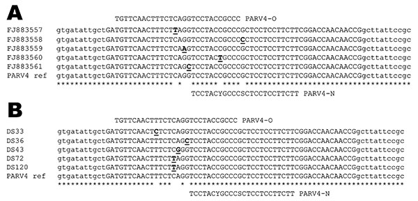
Figure. Alignment of parvovirus 4 (PARV4) sequences showing the location of the 2 probes used in the real-time experiments. A) Partial sequences used for the design of probe PARV4-N: PARV4 prototype isolate (AY622943) and in-house PCR products characterized initially (GenBank accession nos. FJ883557–61). B) Examples of point mutations located on the PARV4-O recognition site identified in amplicons originating from samples positive with PARV4-N assay. Mismatches identified in the alignments are underlined. Nucleotides shown in lowercase letters correspond to 5′/3′ ends of the real-time primers. Mismatches identified in the alignments are underlined and in boldface.
Page created: March 31, 2011
Page updated: March 31, 2011
Page reviewed: March 31, 2011
The conclusions, findings, and opinions expressed by authors contributing to this journal do not necessarily reflect the official position of the U.S. Department of Health and Human Services, the Public Health Service, the Centers for Disease Control and Prevention, or the authors' affiliated institutions. Use of trade names is for identification only and does not imply endorsement by any of the groups named above.