Volume 16, Number 3—March 2010
Dispatch
Sarcocystis Species Lethal for Domestic Pigeons
Philipp Olias , Achim D. Gruber, Andrea Kohls, Hafez M. Hafez, Alfred Otto Heydorn, Heinz Mehlhorn, and Michael Lierz1
Figure 2
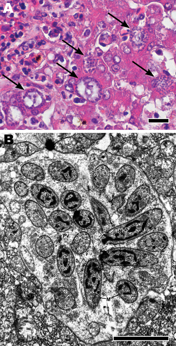
Figure 2. A) Microscopic appearance of liver with tissue necrosis, lymphohistiocytic inflammation, and Sarcocystis schizonts (arrows) in a pigeon 8 days after infection with 105 Sarcocystis sporocysts. Hematoxylin and eosin stain; scale bar = 20 μm. B) Transmission electron micrograph of a hepatocyte from liver in panel A, containing a schizont, forming cross-sectioned and longitudinally sectioned merozoites. N, nucleus; PV, parasitophorous vacuole; M, merozoite. Scale bar = 20 μm.
Page created: December 14, 2010
Page updated: December 14, 2010
Page reviewed: December 14, 2010
The conclusions, findings, and opinions expressed by authors contributing to this journal do not necessarily reflect the official position of the U.S. Department of Health and Human Services, the Public Health Service, the Centers for Disease Control and Prevention, or the authors' affiliated institutions. Use of trade names is for identification only and does not imply endorsement by any of the groups named above.