Volume 17, Number 1—January 2011
Dispatch
Seasonal Influenza A (H1N1) Infection in Early Pregnancy and Second Trimester Fetal Demise
Richard W. Lieberman , Natasha Bagdasarian, Dafydd Thomas, and Cosmas Van De Ven
Figure 1
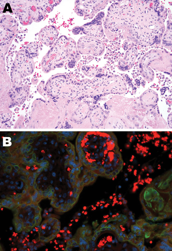
Figure 1. Tissue sample from 30-year-old primigravida patient exposed to seasonal influenza (H1N1). A) Intervillous (maternal) spaces with clusters/sheets of histiocytes (chronic intervillositis) and fibrotic fetal chorionic villi with Hofbauer cells–histiocytic inflammation (hematoxylin and eosin stain, original magnification ×200). B) Dual-stained immunofluorescent assay showing antibodies to influenza A virus (H1N1) (tetramethylrhodamine isothiocyanate, red) and cytokeratin (fluorescein isothiocyanate, green) in intravillous (fetal) and intervillous (maternal) space.
Page created: June 30, 2011
Page updated: June 30, 2011
Page reviewed: June 30, 2011
The conclusions, findings, and opinions expressed by authors contributing to this journal do not necessarily reflect the official position of the U.S. Department of Health and Human Services, the Public Health Service, the Centers for Disease Control and Prevention, or the authors' affiliated institutions. Use of trade names is for identification only and does not imply endorsement by any of the groups named above.