Volume 17, Number 1—January 2011
Dispatch
Seasonal Influenza A (H1N1) Infection in Early Pregnancy and Second Trimester Fetal Demise
Richard W. Lieberman , Natasha Bagdasarian, Dafydd Thomas, and Cosmas Van De Ven
Figure 2
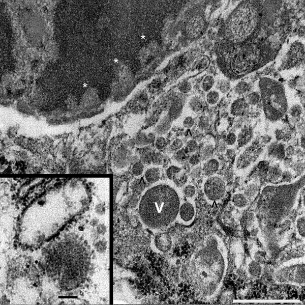
Figure 2. Tissue sample from 30-year-old primigravida patient exposed to seasonal influenza (H1N1). Electron microscopy (original magnification ×70,000) of maternal intervillous space and fetal chorionic villi, showing intranuclear viral transcription aligning along the nuclear envelope–electron hypodensities (asterisks), and intracytoplasmic viral production in varying stages shown by numerous electron densities (V and ^). Scale bar = 500 nm. Inset: enlarged image of mature virion with capsular projections. Ultrastructural features are characteristic of influenza viruses. Scale bar = 100 nm.
Page created: June 30, 2011
Page updated: June 30, 2011
Page reviewed: June 30, 2011
The conclusions, findings, and opinions expressed by authors contributing to this journal do not necessarily reflect the official position of the U.S. Department of Health and Human Services, the Public Health Service, the Centers for Disease Control and Prevention, or the authors' affiliated institutions. Use of trade names is for identification only and does not imply endorsement by any of the groups named above.