Volume 17, Number 10—October 2011
Letter
Sporotrichosis Caused by Sporothrix mexicana, Portugal
Figure
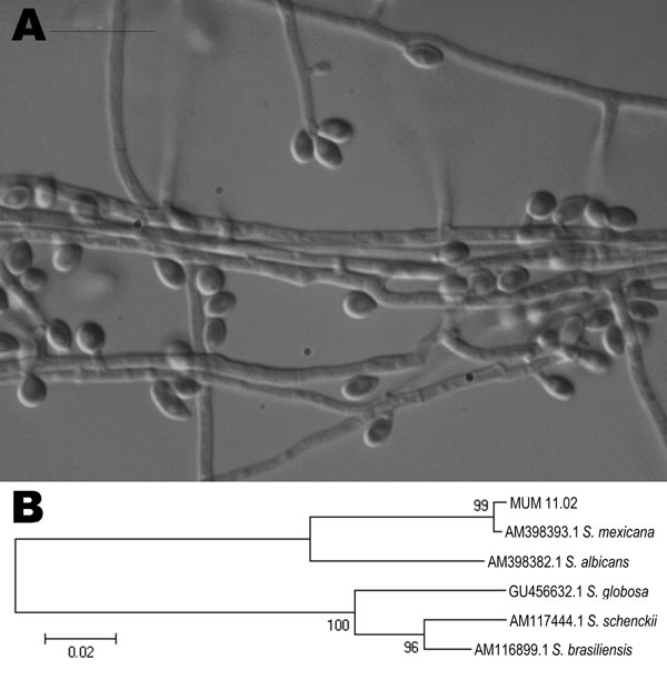
Figure. A) Photomicrograph of sympodial and sessile conidia of Sporothrix mexicana obtained by using a transmitted differential interference contrast microscope. The isolate was obtained from a patient in Portugal in 2009 and archived in the Micoteca da Universidade do Minho (MUM) under accession no. MUM 11.02. Scale bar = 10 μm. B) Neighbor-joining tree showing relatedness of MUM 11.02 isolate with other species of the S. schenckii complex. The percentage of replicate trees in which the associated taxon clustered in the bootstrap test (1,000 replicates) is shown next to the branches. All positions containing gaps and missing data were eliminated from the dataset (complete deletion option). There were 537 positions in the final dataset. Scale bar indicates nucleotide substitutions per site.