Volume 17, Number 11—November 2011
Dispatch
Ultrastructural Characterization of Pandemic (H1N1) 2009 Virus
Cynthia S. Goldsmith , Maureen G. Metcalfe, Dominique C. Rollin, Wun-Ju Shieh, Christopher D. Paddock, Xiyan Xu, and Sherif R. Zaki
Figure 1
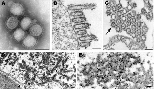
Figure 1. Electron microscopy of pandemic (H1N1) 2009 virus. A) Negatively stained virions grown in MDCK cells showing spherical particles with distinct surface projections. Scale bar = 100 nm. B) Filamentous and ovoid particles assembling at the plasma membrane. Scale bar = 100 nm. C) Extracellular particles showing internal nucleocapsids, seen in cross-section, surrounded by an envelope with prominent spikes. Note all 8 nucleocapsids present in 1 virion (arrow). Scale bar = 100 nm. D) Dense tubules (arrow), which were found in the nuclei of some MDCK-infected cells. Arrowhead, nuclear envelope. Scale bar = 100 nm. E) Immunogold labeling of the nuclear tubules by using an antibody against the matrix protein. Scale bar = 100 nm.
Page created: October 24, 2011
Page updated: October 24, 2011
Page reviewed: October 24, 2011
The conclusions, findings, and opinions expressed by authors contributing to this journal do not necessarily reflect the official position of the U.S. Department of Health and Human Services, the Public Health Service, the Centers for Disease Control and Prevention, or the authors' affiliated institutions. Use of trade names is for identification only and does not imply endorsement by any of the groups named above.