Volume 17, Number 11—November 2011
Dispatch
Ultrastructural Characterization of Pandemic (H1N1) 2009 Virus
Cynthia S. Goldsmith , Maureen G. Metcalfe, Dominique C. Rollin, Wun-Ju Shieh, Christopher D. Paddock, Xiyan Xu, and Sherif R. Zaki
Figure 2
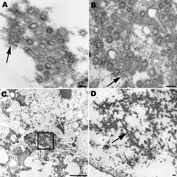
Figure 2. Spherical and ovoid extracellular pandemic (H1N1) 2009 virus particles in human lung tissue found in the alveolar space (A) and in a submucosal gland (B). Nucleocapsids and surface projections are visible on some virions. Note the dense material (arrows) associated with the particles. Scale bars = 100 nm. C) Low-power magnification of the aggregation of virus particles seen in panel B, showing virions (box) in the mucus of the submucosal gland. Scale bar = 1 μm. D) Dense tubules (arrow) found in the nucleus of an infected cell in alveolar space. Scale bar = 100 nm.
Page created: October 24, 2011
Page updated: October 24, 2011
Page reviewed: October 24, 2011
The conclusions, findings, and opinions expressed by authors contributing to this journal do not necessarily reflect the official position of the U.S. Department of Health and Human Services, the Public Health Service, the Centers for Disease Control and Prevention, or the authors' affiliated institutions. Use of trade names is for identification only and does not imply endorsement by any of the groups named above.