Volume 17, Number 4—April 2011
Dispatch
Hepatitis A Virus Vaccine Escape Variants and Potential New Serotype Emergence
Figure 1
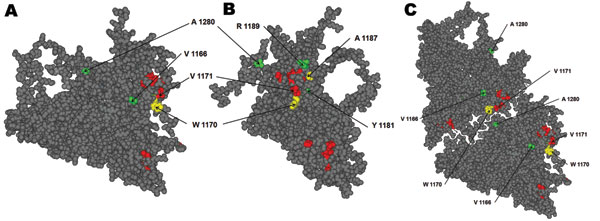
Figure 1. Hepatis A virus protomer model (11; refined by Ming Luo, University of Alabama, Birmingham, AL, USA), which includes the locations of all of the substituted residues in viral protein 1 detected in the isolated variants during 2005–2009. A) Front view of the external surface. B) Lateral view. C) View of 2 adjacent protomers, showing the close contact of residues 1171 and 1280. Red, residues forming the immunodominant site; yellow, residues substituted in monoclonal antibody–resistant mutants C6 (W1170C) and P29 (A1187P); green, residues substituted in the identified natural variants. The amino acid substitution V1171A detected in 1 variant is shown in red because this residue belongs to the immunodominant site.
1These authors contributed equally to this article.