Volume 17, Number 5—May 2011
Historical Review
Evidence of Tungiasis in Pre-Hispanic America
Cite This Article
Citation for Media
Abstract
Ancient parasites of the genus Tunga originated in America and, during the first half of the 19th century, were transported to the Eastern Hemisphere on transatlantic voyages. Although they were first documented by Spanish chroniclers after the arrival of Columbus, little is known about their presence in pre-Hispanic America. To evaluate the antiquity of tungiasis in America, we assessed several kinds of early documentation, including written evidence and pre-Incan earthenware reproductions. We identified 17 written documents and 4 anthropomorphic figures, of which 3 originated from the Chimu culture and 1 from the Maranga culture. Tungiasis has been endemic to Peru for at least 14 centuries. We also identified a pottery fragment during this study. This fragment is the fourth representation of tungiasis in pre-Hispanic America identified and provides explicit evidence of disease endemicity in ancient Peru.
Tungiasis, a parasitic skin disease that occurs in tropical countries, is caused by sand fleas of the genus Tunga (Insecta, Siphonaptera, Tungidae). The disease has recently attracted attention because of high rates of infection for impoverished communities in South America and sub-Saharan Africa and because of new cases reported worldwide as exotic infections among travelers returning to North America and Europe from disease-endemic areas (1,2). The first species, Tunga penetrans, was described by Linnaeus in the 18th century (Pulex penetrans, Linnaeus 1758) (3), and a second Tunga species, which infects humans (T. trimamillata), was taxonomically described by Pampiglione et al. in 2002 (4,5). Studies describing the entomology and pathogenesis of both species are now abundant (6,7). However, studies describing the recognition, proper identification, and distribution of the species in Ecuador and Peru, the only 2 countries where the second new species has been reported, are lacking (8).
Figure 1
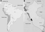
Figure 1. A) Geopolitical map of the Incan Empire at the time of its greatest expansion (dark gray shading). B) Geographic location of the Chimu (dark gray shading) and Maranga (black shading) cultures...
Before the Spanish Conquest, nuclear America was a geopolitical area where the main indigenous populations and cultures were located and where cultural development took place more rapidly than anywhere else in the Americas. It was composed of the 2 centers of the New World, Mesoamerica (Aztecs and Mayas) in the north and the Andean Area (Incas) in the south. The Inca Empire (
Although tungiasis was recognized and documented by Spanish chroniclers shortly after the arrival of Columbus in Central America in 1492 (11), the South American ancestors of the Incas distinguished this affliction from others and depicted it on clay jars, pottery, and ceramics, called huacos in Peru (12–14). Many other autochthonous diseases of ancient Peruvians have also been portrayed on anthropomorphic vessels, thus providing indirect evidence of their presence in this part of the continent (15). Most of this pottery was initially discarded by the Spanish invaders, who looted sacred places, temples, and tombs in their search for gold in the mid-16th century. However, at the turn of the century, interest in pre-Incan cultures and their legacy increased, and these anthropologic pieces represented a cornerstone for understanding the dynamics of cultures that antedated the Incas.
Our objective was to evaluate the antiquity of tungiasis in pre-Hispanic America through the assessment of different kinds of early documentation from 1 of the most advanced civilizations of pre-Hispanic America, which was in Peru. Because documentation of the tungiasis presence in Peru is scarce, we conducted an extensive retrospective search that involved the critical appraisal and inspection of 2 main classes of materials: written evidence and earthenware representations.
During our studies, a pottery fragment was newly identified in a collection storage facility at the Amano Museum Foundation in Lima, Peru. This unique polychromic fragment is the fourth earthenware representation of early tungiasis in Peru identified to date and the only one that represents the different stages of Tunga spp. infection, which distinguishes it from previously described pre-Incan pottery.
To critically inspect written evidence and cover all available information relating to the presence of Tunga spp. in Peru, we searched for all documented names ascribed to this parasite over the past 4 centuries (16–19). We used 35 local terms (nigua, nihua, niua, pique, pigue, piqui, piki, pico, sico, seccec, chegoe, chego, chigger, puce-chique, puce de sable, chique, chica, bicho de pé, bicho do porco, pulga de areia, jatecuba, jigger, chicque, sand flea, tchike, tschike, sike, xique, ckicke, aagrani, atten, tom, tü, tungay, and tunga) and 9 scientific terms (Pulex minimus cutem penetrans Americanus, Pulex minutissimun nigricans, Acarus fuscus sub cutem nidulans proboscide acutiore, Pulex penetrans, Rhynchoprion penetrans, Sarcophaga penetrans, Dermatophilus penetrans, Sarcopsylla penetrans, and Tunga penetrans). Using on-site electronic catalogs, we screened all available manuscripts, books, doctoral theses, journals, bulletins, monographs, and periodicals in their original English, Spanish, or French from 2 major sources: the Main Campus Library of the School of Medicine at Cayetano Heredia Peruvian University, in Lima, Peru, and the William H. Welch Medical Library, Institute of History of Medicine at Johns Hopkins University, in Baltimore, Maryland, USA. These searches were complemented by using the PubMed, LILACS, Scielo, and Medline electronic databases with no publication date- or language-based restrictions. Digitized and printed materials were screened.
After screening the written material to identify the locations of ceramics portraying tungiasis, we assessed earthenware representations through visits to selected private collections of pre-Incan pottery at the Amano Museum Foundation in Miraflores, Lima, Peru, and the Halls of Mexico, Central and South American Peoples at the American Museum of Natural History in New York, New York, USA. These museums were the only facilities cited at least 1 time as potential depositories of pottery depicting pre-Incan tungiasis. All anthropomorphic ceramics that depicted >1 nodule-like representations on the lower or upper extremities, either localized or clustered, with or without representations of holes in the soles of the feet and irrespective of the presence of a central depression, were deemed possible depictions of Tunga spp. infection. From each museum, ≈50 pieces were screened; data on the date and location of findings were recorded when they fulfilled the criteria for possible depiction of tungiasis. A complete screening of the entire collection of ceramics representing diseases of Ancient Peruvians was possible only at the Amano Museum Foundation.
We found written evidence of tungiasis in pre-Incan or Incan times in 17 documents (7 in English, 4 in French, and 6 in Spanish) (Table 1). The documents were 1 unique 17th-century manuscript written by the indigenous Peruvian chronicler Guaman Poma de Ayala (finalized during 1615–1616), 1 monograph, 1 bulletin, 2 doctoral theses, 5 books, and 7 journals. The timeframe in which these documents were written extends from 1615 through 1990.
As for the earthenware representations, we identified 4 anthropomorphic figures representing pre-Incan tungiasis (Table 2). Of these 4 figures, 3 were reproduced in the written materials surveyed (1 from an unknown location and 2 from the American Museum of Natural History), and 1 was a piece of polychromic ceramic, located in the Amano Museum Foundation, which had not been previously described.
Figure 2
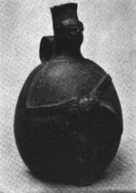
Figure 2. Chimu culture huaco depicting a person extracting parasites with an awl from the sole of the left foot. Multiple holes of various sizes can be seen on the huaco.
The anthropomorphic pottery shown in Figure 2 originated from the Chimu Culture (c.
Figure 3
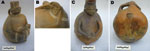
Figure 3. Two globular Chimu huacos found in Pachacamac, a sandy land area in northern Lima. Each person is examining the soles of the feet, on which multiple punch-out lesions can be detected....
The 2 pieces of anthropomorphic pottery shown in Figure 3 also originated from the Chimu culture. They depict 2 men observing the soles of their feet, which show multiple holes of varying sizes. The pieces are located in the American Museum of Natural History but are not on display. They had been found in the Pachacamac Valley, a sandy area in modern southern Lima (Figure 1, panel B).
Figure 4
Figure 4. A) Polychromic Maranga culture fragment that portrays a torso and a tattooed left leg of a person holding a stick while extracting foreign bodies. Cluster lesions with elevated nodules and a...
The anthropomorphic piece shown in Figure 4 originated from the Maranga culture (c.
Tungiasis is an old disease that has been endemic to Peru for centuries and has been illustrated by anthropomorphic pottery showing pathognomonic lesions at various stages of progression. Although the Incas and their ancestors lacked a written language, they used pottery to depict diseases, customs, ceremonies, rituals, and many other activities, thus creating a visual record of their knowledge of a disease process that existed for centuries; such pieces of pottery now provide vivid documentation of their sufferings.
The huaco from Chicama Valley (Figure 2) was described by the Harvard-educated Peruvian archeologist Julio C. Tello (1880–1947). Tello was the first indigenous archeologist of America and is considered the father of Peruvian archeology. In 1924, he reproduced this vessel in a collection of 280 pictures of pottery originating from the Mochica (Moche or Muchik) culture titled Arte Antiguo Peruano, volume II; all pieces depicted in this work are distributed among various museums in Lima (20). Multiple holes in the left plantar surface of the depicted person, a distinguishing feature of tungiasis, can be observed in this figure. Furthermore, the depicted person holds an awl-like instrument in its right hand, which was then commonly used for removing the parasite from the skin. As a physician, Tello easily recognized these lesions as signs of tungiasis or piquinosis (pique infection) (22). However, this collection of pictures of Mochican pottery lacks detailed information about where they were originally found or where they were at the time of its publication. Although it was reproduced as part of a Mochica collection, this huaco actually originates from the Chimu culture. Indeed, the Mochica culture (
A reproduction of the same huaco (Plate 65 in Tello’s Mochica collection) was published 14 years earlier, in 1910, by Albert S. Ashmead (1850–1911) (30). Ashmead was one of the first North American physicians to study Peruvian potteries that depicted diseases, predominantly leprosy and syphilis, at the beginning of the 20th century (32). He received a diverse array of pictures of huacos (including that in Figure 2) directly from Tello, with whom he corresponded regularly. With Tello’s permission, Ashmead subsequently published his reproduction of this huaco (22). Unlike Tello, Ashmead documented the site at which this huaco was originally found, the Chicama Valley (this information was probably provided by the archeologist who discovered the piece). Nevertheless, Ashmead did not associate these lesions with tungiasis and instead thought they were a product of syphilis. In a letter addressed to Ashmead, Tello uses the word piquinosis to describe to the tungiasis depicted on huaco 1; unfortunately, Ashmead did not recognize this regional term (piquinosis = pique infection) used to designate tungiasis (22).
The 2 huacos from the Pachacamac Valley (Figure 3) were first published by Ashmead in 1907 (21). As with the huaco from the Chicama Valley (Figure 2), Ashmead erroneously concluded that the lesions depicted on the soles of the feet of persons depicted on these 2 jars represented signs of uta, which he believed to be skin tuberculosis (the etiology of uta, or cutaneous leishmaniasis, in Peru was later unveiled during the 1913 Harvard expedition to the Amazon region led by Richard P. Strong (33). Because he was interested in prehistoric syphilis and Peruvian earthenware representing diseases, Ashmead maintained correspondence with several renowned physicians from Lima, including Tello. However, Ashmead never associated these 2 huacos with tungiasis, arguing that the holes in the feet were too prominent to represent tungiasis (32). It was paleopathologist Roy L. Moodie (1880–1934) and then Americanist Raoul d’Harcourt (1879–1971) who later reevaluated the significance of these vessels, both concurring that the holes on the feet of these 2 huacos represent residual lesions left by nigua infections (12,23,24,26). Pachacamac, the site at which these 2 jars were located, was not part of the Chimu culture’s territory (Figure 1, panel B). Because the Old Sanctuary of Pachacamac was the major place of worship of the pre-Hispanic Peruvian coast for >1,500 years (31), its temples were visited by masses of pilgrims from the entire Andean world, who carried with them diverse offerings, including huacos, during religious rituals and ceremonies. Thus, archeological pieces from the coastal, highland and Amazon regions of Peru can be found in Pachacamac.
During our visit to the Amano Museum Foundation in 2009, we found the fragment of a huaco from Las Palmas (Figure 4) in a private collection storage room. It had originally been excavated by Yoshitaro Amano (1898–1982), a prosperous Japanese businessman who arrived in Peru in 1951 and was captivated by its history. He excavated and rescued innumerable pieces from sacked and abandoned archeological sites. Pedro Weiss (1893–1985), a Peruvian pathologist who dedicated part of his life to the study of these potteries, mentioned that there were representations of niguas in this museum in his 1980 article La Enfermedad en las Creencias de los Primitivos Americanos; however, he neither photographed nor described any huacos (29). In contrast to the evidence we have for the previously described huacos, we do not have strong evidence proving that this fragment was the one described by Weiss in his above-mentioned work. Together with the first 3 vessels described here, which were also cited by Hoeppli in 1959 as early documentation of parasites in the Western Hemisphere (28), to our knowledge, this fragment is the fourth representation of Tunga spp. infection identified in pre-Hispanic American art. Furthermore, it is the only vessel that depicts different stages of tungiasis, thus representing explicit evidence of its endemicity in ancient Peru.
Along with these 4 huacos, additional evidence suggests the presence of tungiasis in pre-Incan Peru. The 2 most common names attributed to the sand flea in Peru and other countries of South America— nigua and pique—come from the Arawak and Quechua languages, respectively. Indeed, Quechua was the official language of the Incan Empire and is currently the second most commonly spoken language in Peru, after Spanish. Furthermore, the Incas named it seccec from the verb seccen, a Quechua word that means itching (16,17). Currently, it is called huchuy piqui (or huchhuy piqui, according to Lavoveria [20]) or ushtuchi piki by Amerindian communities in the Highlands.
Another aspect of pre-Incan tungiasis is documentation of the therapeutic approaches by historians, anthropologists, and physicians. For example, in his book La Médecine dans l’Ancien Pérou, d’Harcourt mentioned that Peruvian natives used a stick to remove fleas from their feet (26), similar to what is observed on our fragment. In addition, Lastres, in his compendious Historia de la Medicina Peruana, mentioned nigua as being endemic to Peru and described the application of sweet potatoes leaves to the feet to treat infections (27).
Until now, numerous factors have impeded our understanding of the history of tungiasis in Peru. First, the sand flea has been given multiple names by populations living in parasite-endemic areas, making literature searches difficult. Nigua, pique, jigger, chigoe, puce-chique, and tchique are only a few of the many names that have been given to this burrowing flea. Second, it has been taxonomically reclassified multiple times with different names by entomologists over the past 3 centuries (16–19,34). Finally, the high rates of endemicity, along with a relatively uncomplicated clinical course, have made it a disease that is underreported and neglected among physicians in Peru (8).
Our search had some limitations. The dispersed distribution of these Peruvian anthropomorphic pieces in art museums throughout the world made it difficult to document the exact number of pottery pieces that depict tungiasis (35). An unknown number of disease-illustrating huacos remain to be located and investigated. At the beginning of the Spanish Conquest, the conquerors looted religious places in their quest for gold, leaving behind innumerable pieces of pottery made by the Incas and their predecessors. Later, at the beginning of the 20th century, theories about the people of the Americas were propounded along with the study of pre-Hispanic cultures. As a result, sacred places, ceremonial paraphernalia, and other anthropologic pieces in the coast and the Andes were unearthed. These clay pottery pieces were deemed rarities and were highly prized by antiquity collectors. In fact, Ashmead and Tello clearly stated that a large number of Peruvian archeological pieces were highly prized on the black market in their time (36,37). Even today, substantial illicit traffic of ceramics from ancient Peru continues, which has forced the International Council of Museums to include Mochica vessels in the Red List of Latin American Cultural Objects at Risk (38).
Our photograph of the newly identified fragment depicting tungiasis provides additional evidence of tungiasis among ancient Peruvians. The knowledge of this disease in pre-Incan cultures is a valuable legacy that gives a historical insight into the endemicity of this arthropod in South America. These anthropological pieces are now dispersed among numerous museums worldwide. Their identification and analytic evaluation is critical for enhancing our understanding of the history and effects of this flea that continues to affect Peruvians today as it did in pre-Incan times.
Dr Maco is an associate investigator at the Institute of Tropical Medicine Alexander von Humboldt in Lima, Peru. His research interest focuses on neglected intestinal parasites and history of tropical medicine in Peru.
Acknowledgments
We thank Justo Caceres Macedo for his guidance and recognition of the vessels described, Mario Amano Watanabe for giving us complete access to his private collection, Simon Ricarde for accommodating the photography sessions during our visit and for his kind explanations, and Catherine de Beaumont for her comments and critical review of the manuscript.
This work was supported by a private fund granted by the Guadalupe Surgical Clinic in Lima, Peru.
References
- Franck S, Feldmeier H, Heukelback J. Tungiasis: more than an exotic nuisance. Travel Med Infect Dis. 2003;1:159–66. DOIPubMedGoogle Scholar
- Mateos-Rodríguez F, Carranza-Rodríguez C, Pisos-Alamo E, Pérez-Arellano JL. Periungual lesions in a traveler returning from South America [in Spanish.]. Enferm Infecc Microbiol Clin. 2008;26:529–30.PubMedGoogle Scholar
- Linnaeus C. Systema naturae per regna tria naturae, secundum classes, ordines, genera, species, cum characteribus, differentiis, synonymis, locis. 1758. Tomus I. Editio decima, reformata. Holmiae, p. 614.
- Pampiglione S, Trentini M, Fioravanti ML, Onore G, Rivasi F. A new species of Tunga (Insecta, Siphonaptera) from Ecuador. Parassitologia. 2002;44(Suppl 1):127.
- Pampiglione S, Fioravanti ML, Gustinelli A, Onore G, Mantovani B, Luchetti A, Sand flea (Tunga spp.) infections in humans and domestic animals: state of the art. Med Vet Entomol. 2009;23:172–86. DOIPubMedGoogle Scholar
- Luchetti A, Mantovani B, Pampiglione S, Trentini M. Molecular characterization of Tunga trimamillata and T. penetrans (Insecta, Siphonaptera, Tungidae): taxonomy and genetic variability. Parasite. 2005;12:123–9.PubMedGoogle Scholar
- Eisele M, Heukelbach J, Van Marck E, Mehlhorn H, Meckes O, Franck S, Investigation on the biology, epidemiology, pathology and control of Tunga penetrans in Brazil: I. Natural history of tungiasis in man. Parasitol Res. 2003;90:87–99.PubMedGoogle Scholar
- Maco V, Maco VP, Gotuzzo E. An ectopic case of Tunga spp. in Peru. Am J Trop Med Hyg. 2010;82:1076–8. DOIPubMedGoogle Scholar
- Mosley ME. Introduction. In: The Incas and their ancestors. London: Thames & Hudson Ltd; 2005. p. 7–24.
- Bankes G. The Moche culture. In: Moche pottery from Peru. London: British Museum Publications Ltd; 1980. p. 9–13.
- Oviedo y Valdes F. Sumario de la natural historia de la Indias. Toledo: acostas del autor: por industrias del maestre Ramon de Petras. 1526.
- Moodie RL. New observations in paleopathology. Ann Med Hist. 1919;2:241–7.
- Hoeppli R. Ancient times to the middle of the seventeenth century—the knowledge of parasites. In: Parasites and parasitic infections in early medicine and science. Singapore: University of Malaya Press; 1959. p. 1–26.
- Guerra F. Nosologia precolombina. In: La medicina precolombina. Madrid: Instituto de Cooperacion Iberoamericana; 1990. p. 89–114.
- Cabieses F. Diseases and the concept of disease in Ancient Peru. In: Bowers JZ, Purcell EF, editors. Aspects of the history of medicine in Latin America. New York: Independent Publishers Group; 1979. p. 16–53.
- Gage-Lebas LL. Des animaux nuisibles à l’homme et en particulier du Pulex penetrans (chique o nigua). Thèse pour le doctorat en médecine, Faculté de Médecine de Paris. Paris: Imprimerie de Victor Goupy; 1867
- Guyon MJ. Histoire naturelle et médicale de la chique Rhynchoprion penetrans (Oken) insecte parasite des regions tropicales des deux Amériques. Paris: Imprimerie de Madame Veuve Bouchard-Houzard; 1870.
- Baker CF. A revision of American Siphonaptera, or fleas, together with a complete list and bibliography of the group. From the Proceedings of the United States National Museums, vol. XXVII; 1904. p. 365–469.
- Russell H. The chigoes and their allies. In: The flea. London: Cambridge University Press; 1913. p. 74–83.
- Lavoreria DE. El arte de curar entre los antiguos peruanos. Tesis presentada por Daniel Eduardo Lavoreria para optar el grado de Doctor en la Facultad de Medicina. Anales de la Universidad Mayor de San Marcos de Lima, publicados por su rector el Dr. D. Francisco Garcia Calderon, Tomo XXIX, Lima (Peru); 1902.
- Ashmead AS. An ancient Peruvian effigy vase exhibiting disease of the foot. Am Anthropol. 1907;9:738–40. DOIGoogle Scholar
- Ashmead AS. Utosic syphilis and some other things of interest to Paleo-American medicine, as represented on the huacos potteries of old Peru: by Albert S. Ashmead, M.D., of Canadensis, Pennsylvania (fourth part). American Journal of Dermatology and Genito-Urinary Diseases. 1910;14:490–503.
- Moodie RL. Studies in paleopathology: the diseases of the ancient Peruvians and some account of their surgical practices. Surgical Clinics of Chicago. 1920;4:211–31.
- Moodie RL. Diseases of the ancient Peruvians. In: Paleopathology: an introduction to the study of ancient evidences of diseases. Urbana (IL): University of Illinois Press; 1923. p. 485–542.
- Tello JC. Tecnologia y Morfologia. In: Tello JC, editor. Arte antiguo Peruano. Vol. II, primera parte. INCA revista de estudios antropologicos (Organo del Museo de Arquelogia de la Universidad Mayor de San Marcos de Lima). Lima (Peru); 1924. Plate 65.
- d’Harcourt R. Le mal et les guérisseurs. In: La médecine dans l’ancien Pérou. La médecine à travers le temps et l’espace, collection publiée sous la direction du Dr Stéphen-Chauvet. Paris: Librairie Maloine; 1939. p. 15–51.
- Lastres JB. La medicina Incaica—las enfermedades. In: Eguiguren LA, editor. Historia de la medicina Peruana, Volumen I. Lima (Peru): Universidad Nacional Mayor de San Marcos, Publicaciones del Cuarto Centenario; 1951. p. 147–62. Spanish.
- Hoeppli R. Parasitic diseases in Africa and the Western Hemisphere—early documentation and transmission by the slave trade. Acta Trop. 1969;Suppl 10:169–76.
- Weiss P. La enfermedad en las creencias de los primitivos Americanos. Boletin de Lima. 1980;6:28–39.
- Ashmead AS. Utosic syphilis and some other things of interest to Paleo-American medicine, as represented on the huacos potteries of old Peru: by Albert S. Ashmead, M.D., of Canadensis, Pennsylvania (first part). American Journal of Dermatology and Genito-Urinary Diseases. 1910;13:329–43.
- Caceres J. Periodo de los Estados Regionales (900 –1,470 d.C.). In: Enriquez GR, editor. Culturas prehispanicas del Peru: guia de arqueologia Peruana. Lima (Peru); 2009. p. 259–331.
- Death of Albert S. Ashmead. The Medical Fortnightly. 1911;34:144–5.
- Harvard University, Department of Tropical Medicine. Medical report of the Hamilton Rice seventh expedition to the Amazon, in conjunction with the Department of Tropical Medicine of Harvard University, 1924–1925. Boston: Harvard University Press; 1926. p. 313.
- Karsten H. Contribution towards the knowledge of the Rhynchoprion penetrans. Annals and Magazine of Natural History, including Zoology. Botany, and Geology. 1865;15:293–312.
- Bankes G. Chronology of Peruvian pottery. In: Cranstone B, editor. Peruvian pottery. Bucks (UK): Shire Publications Ltd; 1989. p. 11–28.
- Ashmead AS. Germany interested in pre-Columbian pottery of America [correspondence]. St. Louis Medical and Surgical Journal. 1902;82:209.
- Tello JC. Un modelo de escenografía plástica en el arte antiguo Peruano. Wira Kocha. Revista Peruana de Estudios Antropológicos. 1931;1:89–112.
- International Councils of Museums. Red list of Latin American cultural objects at risk [updated 2003 Oct; cited 2010 Mar 16]. http://archives.icom.museum/redlist/LatinAmerica/english/page03.htm
Figures
Tables
Cite This ArticleTable of Contents – Volume 17, Number 5—May 2011
| EID Search Options |
|---|
|
|
|
|
|
|
Please use the form below to submit correspondence to the authors or contact them at the following address:
Vicente Maco, Institute of Tropical Medicine Alexander von Humboldt. Av Honorio Delgado s/n Lima, Lima, Peru
Top