Volume 17, Number 6—June 2011
Letter
Mimivirus-like Particles in Acanthamoebae from Sewage Sludge
William H. Gaze , Gina Morgan, Lihong Zhang, and Elizabeth M.H. Wellington
Figure
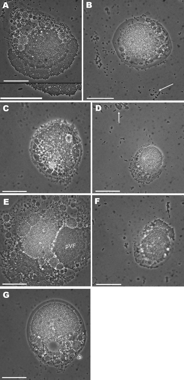
Figure. Light micrograph images of acanthamoebae infected with mimivirus-like particles, showing cells packed with mimivirus-like particles. Enlargement of region of image is shown at bottom of panel A. White arrows in panels B and D indicate free virus-like particles. pVF, putative viral factory. Scale bars = 10 µm.
Page created: August 03, 2011
Page updated: August 03, 2011
Page reviewed: August 03, 2011
The conclusions, findings, and opinions expressed by authors contributing to this journal do not necessarily reflect the official position of the U.S. Department of Health and Human Services, the Public Health Service, the Centers for Disease Control and Prevention, or the authors' affiliated institutions. Use of trade names is for identification only and does not imply endorsement by any of the groups named above.