Volume 17, Number 7—July 2011
CME ACTIVITY - Synopsis
Neurognathostomiasis, a Neglected Parasitosis of the Central Nervous System
Juri Katchanov, Kittisak Sawanyawisuth, Verajit Chotmongkol, and Yukifumi Nawa
Figure 1
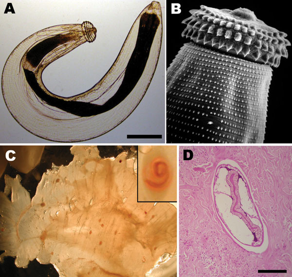
Figure 1. A) Third-stage larva of the nematode Gnathostoma sp. Scale bar = 250 µm. B) Scanning electronic microscopy image depicting head bulb with 4 cephalic hooklet rows. Original magnification ×500. C) Gnathostoma sp. larvae in the flesh of their intermediate host, Eleotris picta fish. Original magnification ×4. Inset: Higher magnification of an encysted larva; original magnification ×100. Larvae photographs courtesy of Dr Diaz-Camacho, Universidad Autónoma de Sinaloa, Sinaloa, Mexico. D) Cross section of a Gnathostoma sp. larva in human skin biopsy sample (hematoxylin and eosin stain). Scale bar = 250 µm.
Page created: August 16, 2011
Page updated: August 16, 2011
Page reviewed: August 16, 2011
The conclusions, findings, and opinions expressed by authors contributing to this journal do not necessarily reflect the official position of the U.S. Department of Health and Human Services, the Public Health Service, the Centers for Disease Control and Prevention, or the authors' affiliated institutions. Use of trade names is for identification only and does not imply endorsement by any of the groups named above.