Skip directly to site content Skip directly to page options Skip directly to A-Z link Skip directly to A-Z link Skip directly to A-Z link
Volume 17, Number 9—September 2011
Dispatch

Tubulinosema sp. Microsporidian Myositis in Immunosuppressed Patient

Maria M. ChoudharyComments to Author , Maureen G. Metcalfe, Kathryn Arrambide, Caryn Bern, Govinda S. Visvesvara, Norman J. Pieniazek, Rebecca D. Bandea, Marlene DeLeon-Carnes, Patricia Adem, Moaz M. Choudhary, Sherif R. Zaki, and Musab U. Saeed
Author affiliations: Author affiliations: Cleveland Clinic Foundation, Cleveland, Ohio, USA (Maria M. Choudhary); Centers for Disease Control and Prevention, Atlanta, Georgia, USA (M.G. Metcalfe, C. Bern, G.S. Visvesvara, N.J. Pieniazek, R.D. Bandea, M. DeLeon-Carnes, P. Adem, S.R. Zaki); Southern Illinois University, Quincy, Illinois, USA (K. Arrambide, Moaz M. Choudhary, M.U. Saeed)

Main Article

Figure 2

Spores of Tubulinosema sp. from a 67-year-old woman with Tubulinosema sp. infection, 2009. A) Electron micrograph of numerous spores in various stages in muscle tissue. Scale bar = 2 μm. B) An immature spore showing an electron-dense exospore (Ex) and a thick electron-lucent endospore (En), which together compose the spore wall. Diplokaryon (DK), posterior vacuole (PV), ribosomes in crystalline clusters as polyribosomes (PR) along with the polaroplast and polar filaments (PF) constitute the spor

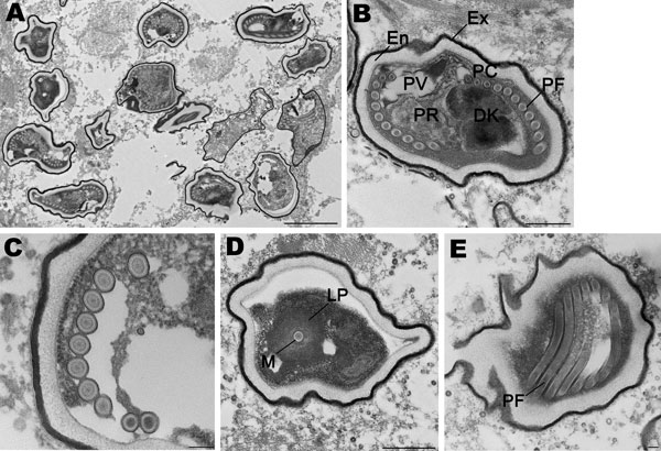
Figure 2. Spores of Tubulinosema sp. from a 67-year-old woman with Tubulinosema sp. infection, 2009. A) Electron micrograph of numerous spores in various stages in muscle tissue. Scale bar = 2 μm. B) An immature spore showing an electron-dense exospore (Ex) and a thick electron-lucent endospore (En), which together compose the spore wall. Diplokaryon (DK), posterior vacuole (PV), ribosomes in crystalline clusters as polyribosomes (PR) along with the polaroplast and polar filaments (PF) constitute the sporoplasm. A small difference in polar filament diameter in the anterior and posterior coils (PC), with the last 3 or 4 coils being smaller (anisofilar), is also apparent. Scale bar = 500 nm. C) Polar filaments exhibiting a lucent ring around a dense core. Scale bar = 100 nm. D) Cross-section of the manubroid (M), which is the straight portion of the polar filament surrounded by the lamellar polaroplast (LP). Scale bar = 500 nm. E) Longitudinal sections of polar filaments (PF) stained with uranyl acetate-lead citrate. Scale bar = 100 nm.

Main Article

Page created: September 06, 2011
Page updated: September 06, 2011
Page reviewed: September 06, 2011
The conclusions, findings, and opinions expressed by authors contributing to this journal do not necessarily reflect the official position of the U.S. Department of Health and Human Services, the Public Health Service, the Centers for Disease Control and Prevention, or the authors' affiliated institutions. Use of trade names is for identification only and does not imply endorsement by any of the groups named above.
file_external