Volume 17, Number 9—September 2011
Dispatch
Tubulinosema sp. Microsporidian Myositis in Immunosuppressed Patient
Figure 2

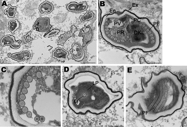
Figure 2. Spores of Tubulinosema sp. from a 67-year-old woman with Tubulinosema sp. infection, 2009. A) Electron micrograph of numerous spores in various stages in muscle tissue. Scale bar = 2 μm. B) An immature spore showing an electron-dense exospore (Ex) and a thick electron-lucent endospore (En), which together compose the spore wall. Diplokaryon (DK), posterior vacuole (PV), ribosomes in crystalline clusters as polyribosomes (PR) along with the polaroplast and polar filaments (PF) constitute the sporoplasm. A small difference in polar filament diameter in the anterior and posterior coils (PC), with the last 3 or 4 coils being smaller (anisofilar), is also apparent. Scale bar = 500 nm. C) Polar filaments exhibiting a lucent ring around a dense core. Scale bar = 100 nm. D) Cross-section of the manubroid (M), which is the straight portion of the polar filament surrounded by the lamellar polaroplast (LP). Scale bar = 500 nm. E) Longitudinal sections of polar filaments (PF) stained with uranyl acetate-lead citrate. Scale bar = 100 nm.