Volume 18, Number 2—February 2012
Letter
Baylisascaris procyonis Infection in Elderly Person, British Columbia, Canada
Tawny Hung, Ronald C. Neafie, and Ian R.A. Mackenzie
Figure
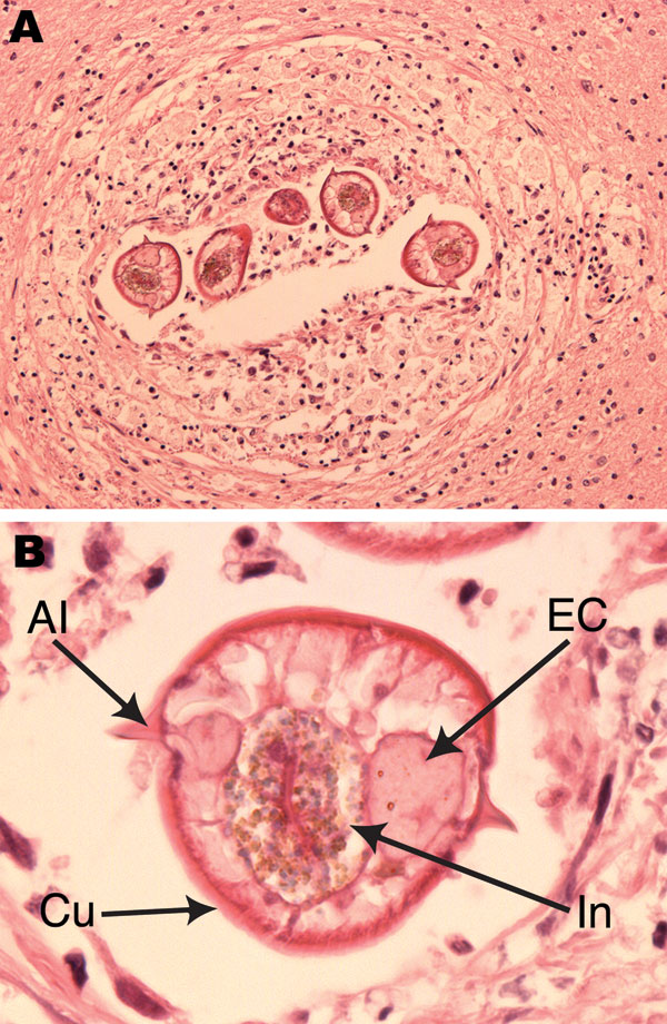
Figure. Baylisascaris procyonis infection in the frontal cerebral lobe white matter. A) Larval nematode seen in multiple transverse sections, surrounded by mild chronic inflammation and reactive changes. Hematoxylin and eosin stain; original magnification ×10. B) Morphologic features of the larvae included maximum diameter of 65 μm; thin, striated cuticle (Cu); single paired lateral alae (Al); and paired excretory columns (EC) that were smaller in diameter than the central intestine (In). Hematoxylin and eosin stain; original magnification ×40.
Page created: January 18, 2012
Page updated: January 18, 2012
Page reviewed: January 18, 2012
The conclusions, findings, and opinions expressed by authors contributing to this journal do not necessarily reflect the official position of the U.S. Department of Health and Human Services, the Public Health Service, the Centers for Disease Control and Prevention, or the authors' affiliated institutions. Use of trade names is for identification only and does not imply endorsement by any of the groups named above.