Volume 18, Number 7—July 2012
Dispatch
Disseminated Microsporidiosis in an Immunosuppressed Patient
Figure 1
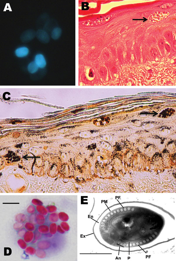
Figure 1. . . . . Microsporidium detected in clinical specimens from a stem cell transplant patient who had undergone substantial immunosuppression. A) Calcofluor white–stained ascitic fluid (original magnification ×500). B) Hematoxylin and eosin–stained skin biopsy sample (original magnification ×400). The arrow indicates clusters of spores. C) Warthin-Starry–stained skin biopsy sample (original magnification ×400). The arrows indicate clusters of spores. D) Modified trichrome–stained material from bronchoalveolar lavage. Scare bar = 5.0 μm. E) Transmission electron micrograph depicting 1 of the microsporidian spores identified in a skin biopsy sample. The image shows the polar filament (PF), containing 13 to 14 coils, in a single layer with anisofilar arrangement (An); the plasma membrane (PM); the exospore (Ex); the endospore (En); and polyribosomes (P). Scale bar = 1 μm.