Volume 18, Number 8—August 2012
Dispatch
Escherichia coli O104 Associated with Human Diarrhea, South Africa, 2004–2011
Cite This Article
Citation for Media
Abstract
To determine the origin of >4,000 suspected diarrheagenic Escherichia coli strains isolated during 2004–2011 in South Africa, we identified 7 isolates as serotype O104; 5 as enteroaggregative E. coli O104:H4, and 2 as enteropathogenic E. coli O104:non-H4. Pulsed-field gel electrophoresis showed that these isolates were unrelated to the 2011 E. coli O104:H4 outbreak strain from Germany.
Escherichia coli is the predominant microorganism of human colonic flora (1). It is mostly harmless to the intestinal lumen. However, some strains (diarrheagenic E. coli [DEC]) can cause disease ranging from moderate-to-severe diarrhea with complications to hemolytic uremic syndrome (1).
During May–June 2011, an outbreak of bloody diarrhea and hemolytic uremic syndrome occurred in Germany and other parts of Europe (2). The Shiga toxin–producing E. coli (STEC) serotype O104 strain was the etiologic agent of this outbreak and accounted for >4,000 cases and 50 deaths (3). This outbreak strain showed an unusual combination of virulence factors of STEC and enteroaggregative E. coli (EAggEC). Furthermore, the outbreak strain showed extended spectrum β-lactamase (ESBL) activity (2,4).
Before the 2011 outbreak, E. coli O104 had been reported in parts of Europe and South Korea (5,6). In South Africa, data for E. coli serotypes are scarce. However, because questions arose about the ancestral origin of the 2011 outbreak strain from Germany, we investigated the occurrence of E. coli O104 associated with human diarrhea in 2 surveillance programs for enteric pathogens in South Africa during 2004–2011. We also investigated phenotypic and genotypic properties of E. coli O104 strains from South Africa and compare these with properties of the outbreak strain from Germany.
The Centre for Enteric Diseases (CED) of the National Institute for Communicable Diseases in South Africa is a reference center for human infections involving enteric pathogens including DEC, Salmonella spp., Shigella spp., and Vibrio cholerae, and it participates in national laboratory-based surveillance for these pathogens. Isolates are voluntarily submitted to the CED from ≈200 clinical microbiology laboratories across the country. The CED also recovers enteric pathogens through its involvement in the Rotavirus Surveillance Project, which started in mid-2009 and involves 5 sentinel hospital sites in South Africa. This project is involved with identification of enteric pathogens (bacterial, viral, and parasitic) associated with diarrhea in children <5 years of age.
All suspected DEC isolates received at the CED were identified by using standard microbiological identification techniques. Serotyping of O antigen was performed by using tube agglutination as described (7). Presence of H4 antigen was determined by PCR detection of the fliCH4 gene (8). Resistance to antimicrobial drugs (ampicillin, amoxicillin/clavulanic acid, sulfamethoxazole/trimethoprim, chloramphenicol, nalidixic acid, ciprofloxacin, tetracycline, kanamycin, imipenem, ceftriaxone, and ceftazidime) was determined by using Etests (bioMérieux, Marcy l’Etoile, France), and ESBL activity was investigated by using double-disk synergy methods, as described by Clinical and Laboratory Standards Institute (Wayne, PA, USA) 2009 guidelines (9).
PCR was used to distinguish DEC strains from nonpathogenic E. coli strains. The PCR consisted of 3 multiplex reactions with published primer sequences. Reactions included 0.2 μmol/L of each primer (Table 1) and 1.5 mmol/L MgCl2. PCR thermal cycling included 35 cycles at 94°C for 1.5 min, 60°C for 1.5 min, and 72°C for 1.5 min.
An isolate was identified as STEC if a PCR result was positive for Shiga toxin genes 1 or 2 (stx1 or stx2); as enterohemorrhagic E. coli if a PCR result was positive for the gene coding intimin outer membrane protein and an stx gene; as enteropathogenic E. coli (EPEC) if a PCR result was positive for the gene coding intimin outer membrane protein; as enterotoxigenic E. coli if a PCR result was a positive for genes coding heat-stable enterotoxin or heat-labile enterotoxin; as enteroinvasive E. coli if a PCR result was positive for the gene coding an invasion protein; as EAggEC if a PCR result was positive for the gene coding a transporter protein; and as diffusely adherent E. coli if a PCR result was positive for the gene coding an accessory protein with a function in F1845 fimbriae production. These genes are listed in Table 1.
Genotypic relatedness of strains was investigated by using a PulseNet protocol (14) for pulsed-field gel electrophoresis (PFGE) of XbaI-digested genomic DNA in a CHEF-DR III electrophoresis system (Bio-Rad Laboratories, Hercules, CA, USA) and the following electrophoresis parameters: voltage 6 V, run temperature 14°C, run time 19 h, initial switch time 6.76 s, final switch time 35.38 s, and included angle 120°. PFGE patterns were analyzed by using BioNumerics version 6.5 software (Applied Maths, Sint-Martens-Latem, Belgium). Dendrograms of patterns were created by using unweighted pair group method with arithmetic averages. Analysis of band patterns incorporated the Dice coefficient at an optimization setting of 1.5% and a position tolerance setting of 1.5%.
During January 2004–May 2011, CED received >4,000 suspected DEC isolates for further laboratory characterization. Of these isolates, 7 (0.2%) were serotype O104. These isolates were collected from Gauteng (n = 3), Mpumalanga (n = 2), and Eastern Cape (n = 1) and North West (n = 1) Provinces of South Africa. E. coli O104 was isolated more often from children <2 years of age than from older children and adults (Table 2). There were more female patients (57%) affected than male patients (43%). Five (71%) of 7 isolates were EAggEC, and 2 (29%) of 7 isolates were EPEC (Table 2). PCR amplification of the fliCH4 gene showed that all EAggEC isolates produced H4 antigen and that all the EPEC isolates did not produce H4 antigen. Therefore, all EAggEC were serotype O104:H4 and all EPEC were serotype O104:non-H4.
EPEC isolates were susceptible to all antimicrobial drugs tested (Table 2). EAggEC isolates were susceptible to amoxicillin/clavulanic acid, chloramphenicol, nalidixic acid, ciprofloxacin, kanamycin, imipenem, ceftriaxone and ceftazidime; were resistant to ampicillin and sulfamethoxazole/trimethoprim; and were variably susceptible to tetracycline and streptomycin (Table 2). None of the EPEC or EAggEC isolates showed ESBL activity.
Figure
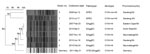
Figure. . Pulsed-field gel electrophoresis profiles (XbaI digestion) of Escherichia coli O104 strains from South Africa (SA) compared with a strain from Germany. EPEC, enteropathogenic E. coli; EaggEC, enteroaggregative E. coli; STEC,...
Dendrogram analysis of PFGE patterns showed that EPEC isolates were diverse and clustered at a pattern similarity of 76%, and that EAggEC isolates were highly clonal and clustered at a pattern similarity of 90% (Figure). The PFGE pattern of the 2011 outbreak strain from Germany did not match those of strains from South Africa. Therefore, the outbreak strain from Germany was determined to be unrelated to strains from South Africa. However, the strain from Germany was most closely related to the EAggEC strain cluster from South Africa (pattern similarity 85%).
Our findings show that E. coli O104 is rarely associated with human diarrhea in South Africa and accounts for <1% of all DEC pathotypes identified during 2004–2011 by laboratory-based surveillance and limited sentinel surveillance. Strains of E. coli O104 from South Africa were mostly associated with EAggEC pathotypes; most (5/7) were identified as EAggEC. Infection with this pathotype has been associated with more persistent diarrhea (duration >14 days) (15). Therefore, patients infected with EAggEC are more likely to have fecal cultures tested, potentially leading to greater numbers of EAggEC isolates identified.
In South Africa, E. coli O104 infections were more commonly identified in children than in adults. Unlike the E. coli O104 strain that caused the outbreak in Germany, strains of E. coli O104 from South Africa did not produce Shiga toxin and did not show ESBL activity. PFGE data supported these phenotypic data, suggesting that strains from South Africa were not related to the outbreak strain from Germany. The PFGE data also showed that strains of EAggEC O104:H4 from South Africa were highly clonal. Further work is necessary to better understand the global distribution of these isolates and the role of molecular epidemiologic techniques in characterizing this newly emerging serotype.
Ms Tau is a medical biological scientist at the Centre for Enteric Diseases, National Institute for Communicable Diseases, a division of the National Health Laboratory Service in Johannesburg, South Africa. Her research interest includes molecular epidemiology of enteric pathogens, in particular Escherichia coli, Vibrio cholerae, Salmonella, and Shigella species, and mechanisms of antimicrobial drug resistance.
Acknowledgments
We thank the Centre for Enteric Diseases, South Africa, for assistance.
This study was supported by the National Institute for Communicable Diseases, a division of the National Health Laboratory Service, South Africa.
References
- Nataro JP, Kaper JB. Diarrheagenic Escherichia coli. Clin Microbiol Rev. 1998;11:142–201.PubMedGoogle Scholar
- Struelens MJ, Palm D, Takkinen J. Enteroaggregative, Shiga toxin–producing Escherichia coli O104:H4 outbreak: new microbiological findings boost coordinated investigations by European public health laboratories. Euro Surveill. 2011;16:pii:19890. PubMedGoogle Scholar
- World Health Organization Regional Office for Europe. International health regulations. Outbreaks of E. coli O104:H4 infection: update 30, 2011 [cited 2012 Apr 10]. http://www.euro.who.int/en/what-we-do/health-topics/emergencies/international-health-regulations/news/news/2011/07/outbreaks-of-e.-coli-o104h4-infection-update-30
- Rubino S, Cappuccinelli P, Kelvin DJ. Escherichia coli (STEC) serotype O104 outbreak causing haemolytic syndrome (HUS) in Germany and France. J Infect Dev Ctries. 2011;5:437–40. DOIPubMedGoogle Scholar
- Mellmann A, Bielaszewska M, Kock R, Friedrich AW, Fruth A, Middendorf B, Analysis of collection of hemolytic uremic syndrome-associated enterohemorrhagic Escherichia coli. Emerg Infect Dis. 2008;14:1287–90. DOIPubMedGoogle Scholar
- Scheutz F, Nielsen EM, Frimodt-Moller J, Boisen N, Morabito S, Tozzoli R, Characteristics of the enteroaggregative Shiga toxin/verotoxin–producing Escherichia coli O104:H4 strain causing the outbreak of haemolytic uraemic syndrome in Germany, May to June 2011. Euro Surveill. 2011;16:pii:19889. PubMedGoogle Scholar
- Orskov I, Orskov F, Jann B, Jann K. Serology, chemistry, and genetics of O and K antigens of Escherichia coli. Bacteriol Rev. 1977;41:667–710.PubMedGoogle Scholar
- European Union Reference Laboratory for. E. coli, Department of Veterinary Public Health and Food Safety. Detection and identification of verocytotoxin–producing Escherichia coli (VTEC) O104:H4 in food by real time PCR, 2011 [cited 2012 Apr 10]. http://www.iss.it/binary/vtec/cont/Lab_Proc_VTEC_O104.pdf
- Clinical and Laboratory Standards Institute. Methods for dilution antimicrobial susceptibility tests for bacteria that grow aerobically. Approved standard, 8th ed. M07–A8. Wayne (PA): The Institute; 2009.
- Cebula TA, Payne WL, Feng P. Simultaneous identification of strains of Escherichia coli serotype O157:H7 and their Shiga-like toxin type by mismatch amplification mutation assay-multiplex PCR. J Clin Microbiol. 1995;33:248–50.PubMedGoogle Scholar
- Vidal M, Kruger E, Duran C, Lagos R, Levine M, Prado V, Single multiplex PCR assay to identify simultaneously the six categories of diarrheagenic Escherichia coli associated with enteric infections. J Clin Microbiol. 2005;43:5362–5. DOIPubMedGoogle Scholar
- López-Saucedo C, Cerna JF, Villegas-Sepulveda N, Thompson R, Velazquez FR, Torres J, Single multiplex polymerase chain reaction to detect diverse loci associated with diarrheagenic Escherichia coli. Emerg Infect Dis. 2003;9:127–31. DOIPubMedGoogle Scholar
- Schmidt H, Knop C, Franke S, Aleksic S, Heesemann J, Karch H. Development of PCR for screening of enteroaggregative Escherichia coli. J Clin Microbiol. 1995;33:701–5.PubMedGoogle Scholar
- Ribot EM, Fair MA, Gautom R, Cameron DN, Hunter SB, Swaminathan B, Standardization of pulsed-field gel electrophoresis protocols for the subtyping of Escherichia coli O157:H7, Salmonella, and Shigella for PulseNet. Foodborne Pathog Dis. 2006;3:59–67. DOIPubMedGoogle Scholar
- Law D, Chart H. Enteroaggregative Escherichia coli. J Appl Microbiol. 1998;84:685–97. DOIPubMedGoogle Scholar
Figure
Tables
Cite This ArticleTable of Contents – Volume 18, Number 8—August 2012
| EID Search Options |
|---|
|
|
|
|
|
|
Please use the form below to submit correspondence to the authors or contact them at the following address:
Nomsa P. Tau, Centre for Enteric Diseases, National Institute for Communicable Diseases, Private Bag X4, Sandringham, 2131, South Africa
Top