Volume 19, Number 1—January 2013
Research
Novel Polyomavirus associated with Brain Tumors in Free-Ranging Raccoons, Western United States
Figure 1
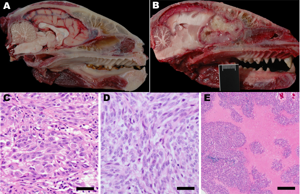
Figure 1. . Pathology of raccoon polyomavirus–associated tumors. A) Normal anatomy, head of unaffected raccoon, midsagittal section. An intact cribriform plate separates the ethmoid turbinates from the olfactory tract. B) Gross pathology, head of raccoon no. 9 (Rac9), left parasagittal section. The tumor obliterates the left olfactory tract and extends into the left frontal lobe to the level of the midbrain. The tumor compresses the brain and distorts the cerebellum. The raccoon head in length (crown to nose) is 16 cm. C) Histopathology (hematoxylin and eosin staining) for Rac5. Marked anisokaryosis and anisocytosis are evident. Original magnification ×40. D) Histopathology (hematoxylin and eosin staining) for Rac10. Shown is a region that has streams of elongated cells. Original magnification ×40. Scale bar = 40 μm. E) Histopathology (hematoxylin and eosin staining) for Rac3. Cell-dense sheets of neoplastic cells are interrupted by vast regions of necrosis. Original magnification ×20.