Volume 19, Number 11—November 2013
Letter
Subcutaneous Infection with Dirofilaria spp. Nematode in Human, France
Figure

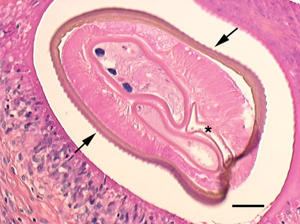
Figure. . Cross-section of the filarial nematode seen in the subcutaneous nodule on the thigh of a woman in France. The features, as described in the original report (1), include prominent, longitudinal ridging of the cuticle (arrows), 2 reproductive tubes, and the intestine (asterisk). Scale bar indicates 50 µm. Image courtesy of Jean-Philippe Dales.
References
- Foissac M, Million M, Mary C, Dales JP, Souraud JB, Piarroux R, Subcutaneous infection with Dirofilaria immitis nematode in human, France. Emerg Infect Dis. 2013;19:171–2 . DOIPubMedGoogle Scholar
Page created: October 31, 2013
Page updated: October 31, 2013
Page reviewed: October 31, 2013
The conclusions, findings, and opinions expressed by authors contributing to this journal do not necessarily reflect the official position of the U.S. Department of Health and Human Services, the Public Health Service, the Centers for Disease Control and Prevention, or the authors' affiliated institutions. Use of trade names is for identification only and does not imply endorsement by any of the groups named above.