Volume 19, Number 12—December 2013
Letter
Concurrent Parasitic Infections in a Renal Transplant Patient
Figure
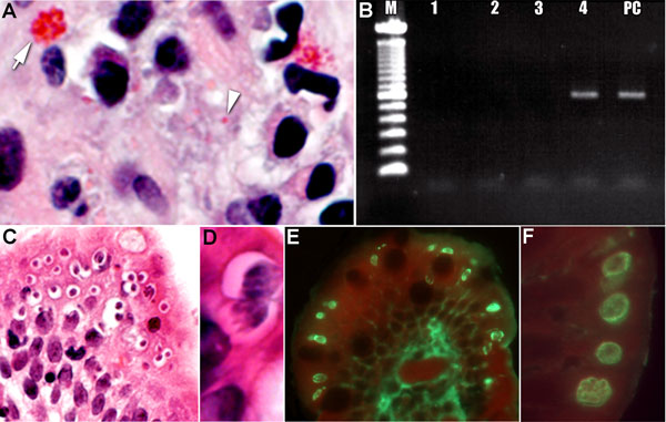
Figure. . Tissue specimens from a kidney transplant recipient with concurrent parasitic infections after traveling to the Dominican Republic. A) Tissue section stained with Gram chromotrope. Note the apical location of a cluster of Enterocytozoon bieneusi spores at arrow (original magnification ×1,000) and single spore at arrowhead.; B) Agarose gel showing PCR amplification of E. bieneusi 18S rDNA in the scraped section, as in panel A (M, 100-bp ladder; lane 1, DNA lysate diluted 1:5; lane 2, 1:10, lane 3, 1: 50 and lane 4, 1:100 of DNA lysate; lane 5 PC, positive control specimen). C) Tissue section stained with hematoxylin and eosin, demonstrating numerous sites in which Cyclospora spores are in developing stages (original magnification ×100). D) Higher power image of Cyclospora spores, showing the developing meronts (original magnification ×1,200). E) Immunofluorescent reactivity (bright green) of the various life cycle stages of Cyclospora with a positive anti-Cyclospora serum sample (original magnification ×200). F) Note the bright fluorescence of the various parasite stages just below the apical (luminal) surface of the epithelial cells (original magnification ×1,000).