Skip directly to site content Skip directly to page options Skip directly to A-Z link Skip directly to A-Z link Skip directly to A-Z link
Volume 19, Number 12—December 2013
Letter

Concurrent Parasitic Infections in a Renal Transplant Patient

Govinda S. Visvesvara, Michael J. ArrowoodComments to Author , Yvonne Qvarnstrom, Rama Sriram, Rebecca Bandea, Patricia P. Wilkins, Eileen Farnon, and Gill Weitzman
Author affiliations: Centers for Disease Control and Prevention, Atlanta, Georgia, USA (G.S. Visvesvara, M.J. Arrowood, Y. Qvarnstrom, R. Sriram, R. Bandea, P.P. Wilkins, E. Farnon); NewYork–Presbyterian/Weill Cornell Medical Center, New York, New York, USA (G. Weitzman)

Main Article

Figure

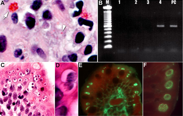
Tissue specimens from a kidney transplant recipient with concurrent parasitic infections after traveling to the Dominican Republic. A) Tissue section stained with Gram chromotrope. Note the apical location of a cluster of Enterocytozoon bieneusi spores at arrow (original magnification ×1,000) and single spore at arrowhead.; B) Agarose gel showing PCR amplification of E. bieneusi 18S rDNA in the scraped section, as in panel A (M, 100-bp ladder; lane 1, DNA lysate diluted 1:5; lane 2, 1:10, lane 3

Figure. . Tissue specimens from a kidney transplant recipient with concurrent parasitic infections after traveling to the Dominican Republic. A) Tissue section stained with Gram chromotrope. Note the apical location of a cluster of Enterocytozoon bieneusi spores at arrow (original magnification ×1,000) and single spore at arrowhead.; B) Agarose gel showing PCR amplification of E. bieneusi 18S rDNA in the scraped section, as in panel A (M, 100-bp ladder; lane 1, DNA lysate diluted 1:5; lane 2, 1:10, lane 3, 1: 50 and lane 4, 1:100 of DNA lysate; lane 5 PC, positive control specimen). C) Tissue section stained with hematoxylin and eosin, demonstrating numerous sites in which Cyclospora spores are in developing stages (original magnification ×100). D) Higher power image of Cyclospora spores, showing the developing meronts (original magnification ×1,200). E) Immunofluorescent reactivity (bright green) of the various life cycle stages of Cyclospora with a positive anti-Cyclospora serum sample (original magnification ×200). F) Note the bright fluorescence of the various parasite stages just below the apical (luminal) surface of the epithelial cells (original magnification ×1,000).

Main Article

Page created: November 20, 2013
Page updated: November 20, 2013
Page reviewed: November 20, 2013
The conclusions, findings, and opinions expressed by authors contributing to this journal do not necessarily reflect the official position of the U.S. Department of Health and Human Services, the Public Health Service, the Centers for Disease Control and Prevention, or the authors' affiliated institutions. Use of trade names is for identification only and does not imply endorsement by any of the groups named above.
file_external