Volume 19, Number 12—December 2013
Dispatch
Cerebellar Cysticercosis Caused by Larval Taenia crassiceps Tapeworm in Immunocompetent Woman, Germany
Figure 1
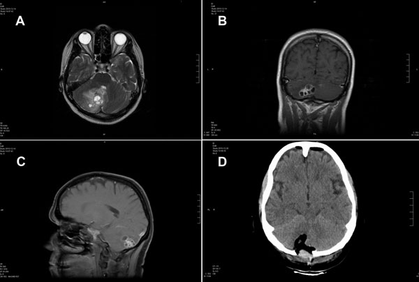
Figure 1. . Magnetic resonance (MR) and computed tomographic images of the brain of a 51-year-old woman infected with Taenia crassiceps tapeworm larvae, Germany. A) Transverse view, T1-weighted MR image. The 30 × 30 mm parasitic lesion with perifocal edema is located in the right hemisphere of the cerebellum and caused ataxia, headache, and nausea. The fourth ventricle is compressed. B) Coronal view, T2-weighted MR image. The cyst-like appearance of the parasitic tissue is clearly visible. This lesion can be misinterpreted as cerebral echinococcosis, racemose cysticercosis caused by a Taenia solium tapeworm, or coenurosis. C) Sagittal view, MR image with contrast enhancing agent. D) Transverse view, computed tomographic image after surgery. No residual parasitic masses, only the parenchymal defect in the cerebellum after resection of T. crassiceps tapeworm larvae, are visible.
1These authors contributed equally to this article.