Volume 19, Number 2—February 2013
Letter
Coxiella burnetii in Ticks, Argentina
Richard C. Pacheco, Ignacio E. Echaide, Rosiane N. Alves, Marcelo E. Beletti, Santiago Nava, and Marcelo B. Labruna
Figure
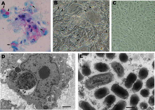
Figure. . Vero cells inoculated with Amblyomma tick extracts for isolation of rickettsiae. A) Rickettsiae-like organisms stained in red by Gimenez staining (original magnification ×400). B) Inoculated monolayer photographed under phase-contrast microscopy (original magnification ×400). C) Uninfected control monolayer under phase-contrast microscopy (original magnification ×400). D) Transmission electron microscopy of infected cells. Bar indicates 2 µm. N, nucleolus. E) Transmission electron microscopy image of intravacuolar bacteria. Bar indicates 250 nm. Arrows indicate vacuoles containing bacteria.
Page created: January 22, 2013
Page updated: January 22, 2013
Page reviewed: January 22, 2013
The conclusions, findings, and opinions expressed by authors contributing to this journal do not necessarily reflect the official position of the U.S. Department of Health and Human Services, the Public Health Service, the Centers for Disease Control and Prevention, or the authors' affiliated institutions. Use of trade names is for identification only and does not imply endorsement by any of the groups named above.