Volume 19, Number 2—February 2013
Dispatch
Hepatitis E Virus in Pork Liver Sausage, France
Alessandra Berto , Sylvia Grierson, Renate Hakze-van der Honing, Francesca Martelli, Reimar Johne, Jochen Reetz, Rainer G. Ulrich, Nicole Pavio, Wim H.M. Van der Poel, and Malcolm Banks
Figure 2
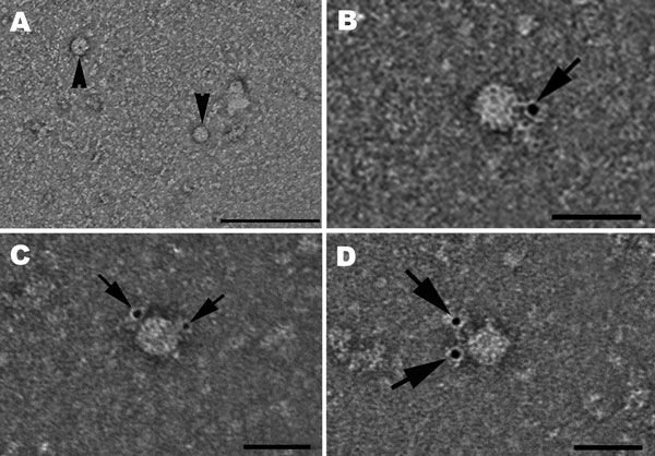
Figure 2. . Hepatitis E virus (HEV) particles in the cell culture supernatant of pork liver sausage sample A, collected at 33 dpi. A) Transmission electron micrograph of negatively stained HEV particles ≈33 and 34 nm (arrowheads). Scale bar indicates 200 nm. B–D) Hepatitis E virions ≈28 (B), 33 (C), or 32 (D) nm in diameter, identified by using an HEV genotype 3–specific rabbit hyperimmune serum and a gold-labeled secondary antibody. Arrows show bound gold particles. Scale bars indicate 50 nm.
Page created: January 22, 2013
Page updated: January 22, 2013
Page reviewed: January 22, 2013
The conclusions, findings, and opinions expressed by authors contributing to this journal do not necessarily reflect the official position of the U.S. Department of Health and Human Services, the Public Health Service, the Centers for Disease Control and Prevention, or the authors' affiliated institutions. Use of trade names is for identification only and does not imply endorsement by any of the groups named above.