Volume 19, Number 3—March 2013
Research
Lack of Norovirus Replication and Histo-Blood Group Antigen Expression in 3-Dimensional Intestinal Epithelial Cells
Melissa M. Herbst-Kralovetz , Andrea L. Radtke, Margarita K. Lay, Brooke E. Hjelm, Alice N. Bolick, Shameema S. Sarker, Robert L. Atmar, David H. Kingsley, Charles J. Arntzen, Mary K. Estes, and Cheryl A. Nickerson
Figure 1
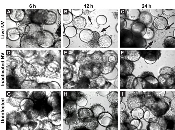
Figure 1. . . Morphologic changes in Norwalk virus (NV)–infected 3-dimensional INT-407 aggregates. Phase contrast micrographs of 3-dimensional intestinal aggregates cultured in the rotating wall vessel bioreactor and subsequently inoculated with live NV (panels A–C), inactivated NV (γ-inactivated) (panels D–F), or phosphate-buffered saline (mock-uninfected control) (panels G–I) at 6, 12, and 24 h after inoculation. Arrows indicate cells (or cellular debris) that were released from the support beads. The beads appear as large, distinct spheres after the removal of bound cells. Original magnification ×20.
Page created: February 12, 2013
Page updated: February 12, 2013
Page reviewed: February 12, 2013
The conclusions, findings, and opinions expressed by authors contributing to this journal do not necessarily reflect the official position of the U.S. Department of Health and Human Services, the Public Health Service, the Centers for Disease Control and Prevention, or the authors' affiliated institutions. Use of trade names is for identification only and does not imply endorsement by any of the groups named above.