Volume 19, Number 7—July 2013
Letter
Human Infection with Marten Tapeworm
Figure
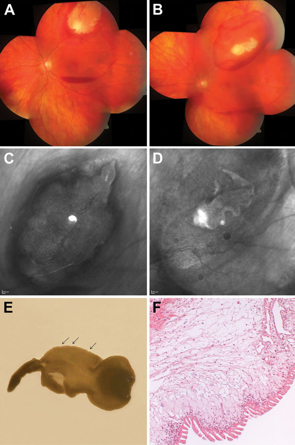
Figure. . . Cysticercosis-like eye infection caused by the tapeworm Taenia martis in a woman. A) Fundus at the patient’s initial visit, before medical therapy. The cyst lies subretinally at the temporal upper branch vessels; adjacent intraretinal and subretinal bleeding and central subhyaloid bleeding can be seen. B) After 8 days of medical therapy, the cyst size had decreased markedly. The physis of the larva (A and B) is reminiscent of the armatetrathyridium (or fimbriocercus), a larval form typical for the tapeworm subspecies T. martis martis. C) Cyst at patient’s initial visit. D) Cyst at the time of surgery. E) Surgically removed monocephalic cysticercus-like larva with inverted parenchymatous portion, withdrawn scolex, and attenuated posterior end. The tegumental surface is transversely striated and exhibits inward folds (arrows). F) Histologic section of the Taenia martis tapeworm cyst showing morphologic characteristics also commonly seen in cysticercosis cysts caused by T. solium tapeworms. The syncytial bladder wall consists of a rugate external, a nucleated intermediate, and an internal reticular layer with lacunate branches of the excretory duct system. Filamentous extensions of contractile muscles project into the parenchyma, which is interspersed with a few calcareous corpuscles. In addition, the T. martis cyst shows a preponderance of uniformly organized, elongate and slender tegumental processes, which are usually not seen in histologic sections of cyst walls caused by T. solium tapeworms. Hematoxylin and eosin stain; objective magnification ×10.