Volume 19, Number 9—September 2013
Research
Divergent Astrovirus Associated with Neurologic Disease in Cattle
Linlin Li, Santiago Diab, Sabrina McGraw, Bradd Barr, Ryan Traslavina, Robert Higgins, Tom Talbot, Pat Blanchard, Guillermo Rimoldi, Elizabeth Fahsbender, Brady Page, Tung Gia Phan, Chunlin Wang, Xutao Deng, Patricia Pesavento , and Eric Delwart
Figure 2
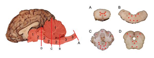
Figure 2. . Yearling steer with encephalomyelitis. Midsagittal section of brain and multiple transverse sections of cerebellum, brainstem, and spinal cord depicting the location and severity of microscopic lesions. Midsagittal section of the brain: red highlight indicates areas of the central nervous system affected, numbers indicate severity of the lesions (1 = least severe; 2 = more severe; 3 = most severe), and red lines (A, B, C, D) indicate the levels where transverse sections were cut. A, spinal cord. B, medulla oblongata. C, cerebellum and cerebellar peduncles. D, midbrain (superior colliculus). Cross-sections of brainstem, cerebellum, and spinal cord: red dots indicate sites and relative intensity of microscopic lesions.
Page created: August 20, 2013
Page updated: August 20, 2013
Page reviewed: August 20, 2013
The conclusions, findings, and opinions expressed by authors contributing to this journal do not necessarily reflect the official position of the U.S. Department of Health and Human Services, the Public Health Service, the Centers for Disease Control and Prevention, or the authors' affiliated institutions. Use of trade names is for identification only and does not imply endorsement by any of the groups named above.