Volume 20, Number 1—January 2014
Dispatch
Fatal Metacestode Infection in Bornean Orangutan Caused by Unknown Versteria Species
Figure 1

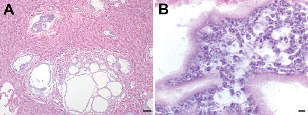
Figure 1. . Microscopic images of liver sections from a Bornean orangutan fatally infected with Versteria metacestodes. Images of liver sections stained with hematoxylin and eosin (H&E) stain were captured at 10× magnification (A; scale bar = 30 μm) and 100× magnification; B; scale bar = 5 μm). Large numbers of parasite cells can be seen within well-defined cystic structures separated from the surrounding host tissue by clearly visible membranes.
Page created: January 03, 2014
Page updated: January 03, 2014
Page reviewed: January 03, 2014
The conclusions, findings, and opinions expressed by authors contributing to this journal do not necessarily reflect the official position of the U.S. Department of Health and Human Services, the Public Health Service, the Centers for Disease Control and Prevention, or the authors' affiliated institutions. Use of trade names is for identification only and does not imply endorsement by any of the groups named above.