Volume 20, Number 12—December 2014
Dispatch
Human Metapneumovirus Infection in Chimpanzees, United States
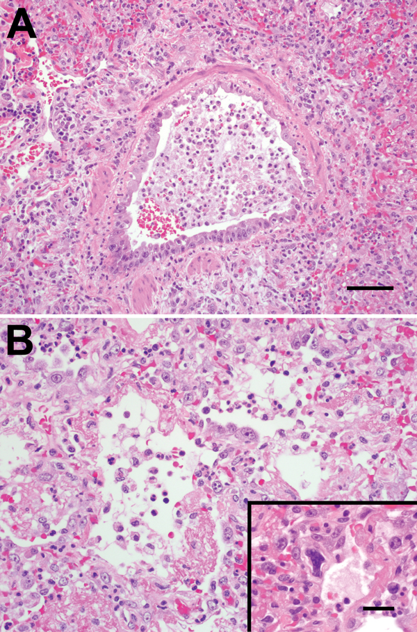
Figure 2

Figure 2. A) Bronchiolar epithelium of chimpanzees infected with human metapneumovirus, United States, 2009, showing cell variation from attenuated to piled and disorganized. Epithelial cells lack cilia, and lumens contain foamy macrophages, neutrophils, and hemorrhage. Adjacent air spaces are filled with similar inflammatory cells. Scale bar = 70 μm. B) Alveoli lined with plump type II pneumocytes and fibrin. Inset: Rare, deeply basophilic, smudged nuclei are present in some areas. Scale bar = 20 μm. Hematoxylin and eosin stain.
1Current affiliation: University of Calgary, Calgary, Alberta, Canada.
Page created: November 19, 2014
Page updated: November 19, 2014
Page reviewed: November 19, 2014
The conclusions, findings, and opinions expressed by authors contributing to this journal do not necessarily reflect the official position of the U.S. Department of Health and Human Services, the Public Health Service, the Centers for Disease Control and Prevention, or the authors' affiliated institutions. Use of trade names is for identification only and does not imply endorsement by any of the groups named above.