Volume 20, Number 12—December 2014
Dispatch
Echinococcus ortleppi Infections in Humans and Cattle, France
Frédéric Grenouillet1 , Gérald Umhang1, Francine Arbez-Gindre, Georges Mantion, Eric Delabrousse, Laurence Millon, and Franck Boué
Figure 1
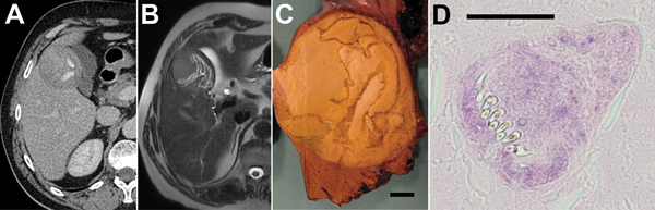
Figure 1. Results of testing in a 63-year-old man from the Jura département (eastern France), who was diagnosed with infection with Echinococcus ortleppi larval tapeworms in 2001. A) Abdominal computed tomography scan; B) magnetic resonance imaging; and C) macroscopic morphologic examination of operative specimen. All show lesions with a detached endocyst and calcified matrix; scale bar in panel C indicates 1 cm. D) Microscopic examination shows evidence of protoscoleces in the matrix (hematoxylin and eosin stain; scale bar indicates 50 µm).
1These authors contributed equally to this article.
Page created: November 19, 2014
Page updated: November 19, 2014
Page reviewed: November 19, 2014
The conclusions, findings, and opinions expressed by authors contributing to this journal do not necessarily reflect the official position of the U.S. Department of Health and Human Services, the Public Health Service, the Centers for Disease Control and Prevention, or the authors' affiliated institutions. Use of trade names is for identification only and does not imply endorsement by any of the groups named above.