Volume 21, Number 1—January 2015
Letter
Serologic Assessment of Possibility for MERS-CoV Infection in Equids
Figure
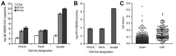
Figure. Analysis of the replication of Middle East respiratory syndrome coronavirus (MERS-CoV) in primary horse kidney cell lines and origin of equine serum samples. A, B) Cells were seeded at densities of 2 × 10⁵ cells/mL and infected in triplicate with a multiplicity of infection of 0.5 infectious MERS-CoV units/cell. After incubation for 1 h, cells were washed twice and supernatants were harvested at 0, 20, and 40 h postinfection (hpi). The replication level is given as the log of the genome equivalents (A) or as PFUs (B). Error bars indicate ranges; PF-N and PFN-R indicate the 2 horse cell lines; VeroB4 is an interferon-deficient primate cell line. C) Distribution of optical density (OD) values (450 nm) of equine serum samples originating from Spain or the United Arab Emirates (UAE).