Volume 21, Number 3—March 2015
Letter
Echinococcus vogeli in Immigrant from Suriname to the Netherlands
Figure
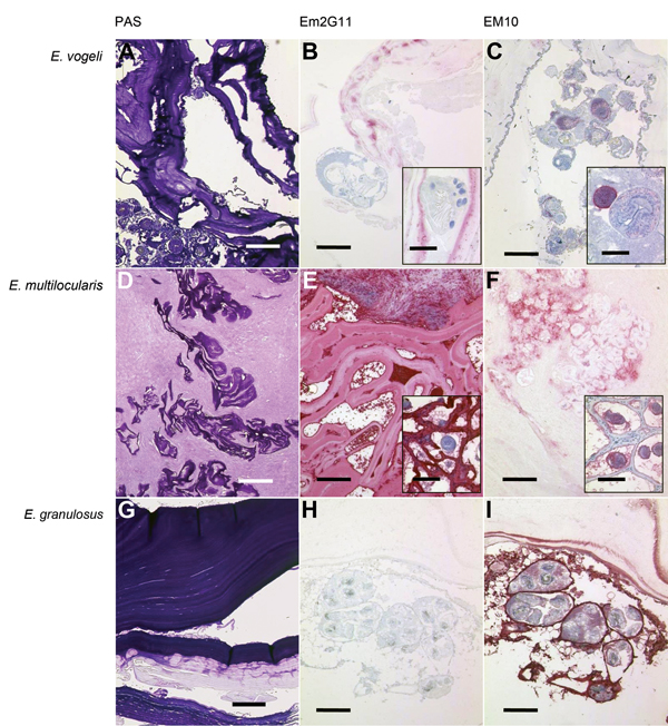
Figure. Periodic acid–Schiff (PAS) staining and immunohistochemical analysis of samples from Echinococcus vogeli lesions, with monoclonal antibodies against Em2 and EM10 of E. vogeli (A, B, C), E. multilocularis (D, E, F), and E. granulosus (G, H, I) lesions. Staining was performed on archived tissue from human patients with alveolar and cystic echinococcosis for comparison, and from the patient with E. vogeli infection who immigrated to the Netherlands from Suriname (E. vogeli infection in 2009). B and C insets) Protoscolex, with rostellar hooks clearly visible in inset B. E and F insets) Tissue from infected rodents (laboratory-infected Meriones unguiculatus gerbils) because E. multilocularis lesions in humans only rarely contain protoscolices. A, D, G) PAS-stained sections of the respective echinococcal lesions. Scale bars indicate 500 μm. B, E, H) Lesions with E. vogeli, E. multilocularis, and E. granulosus infection, respectively, stained with the monoclonal Em2G11 antibody against Em2 (for staining details see [7]). E. multilocularis lesions show intense staining, E. granulosus lesions show no staining, and E. vogeli lesions show patchy stains. Scale bars indicate 200 μm; scale bars of the insets indicate 50 μm. C, F, I) Respective lesions stained with antibodies against EM10 (dilution of the primary antibody 1:50; further steps as in Barth et al. [7]). Germinal layer and protoscolices of E. multilocularis and E. granulosus larvae are stained, but the protoscolices of the E. vogeli metacestode are only partly stained. Scale bars indicate 200 μm; scale bars of the insets indicate 50 μm.
References
- D’Alessandro A, Rausch RL. New aspects of neotropical polycystic (Echinococcus vogeli) and unicystic (Echinococcus oligarthrus) echinococcosis. Clin Microbiol Rev. 2008;21:380–401. DOIPubMedGoogle Scholar
- Tappe D, Stich A, Frosch M. Emergence of polycystic neotropical echinococcosis. Emerg Infect Dis. 2008;14:292–7. DOIPubMedGoogle Scholar
- Knapp J, Chirica M, Simonnet C, Grenouillet F, Bart JM, Sako Y, Echinococcus vogeli infection in a hunter, French Guiana. Emerg Infect Dis. 2009;15:2029–31. DOIPubMedGoogle Scholar
- Stijnis C, Bart A, Brosens L, Van Gool T, Grobusch M, van Gulik T, First case of Echinococcus vogeli infection imported to the Netherlands, January 2013. Euro Surveill. 2013;18:20448 .PubMedGoogle Scholar
- Bowles J, Blair D, McManus DP. Genetic variants within the genus Echinococcus identified by mitochondrial DNA sequencing. Mol Biochem Parasitol. 1992;54:165–73. DOIPubMedGoogle Scholar
- Deplazes P, Gottstein B. A monoclonal antibody against Echinococcus multilocularis Em2 antigen. Parasitology. 1991;103:41–9 . DOIPubMedGoogle Scholar
- Barth TF, Herrmann TS, Tappe D, Stark L, Gruner B, Buttenschoen K, Sensitive and specific immunohistochemical diagnosis of human alveolar echinococcosis with the monoclonal antibody Em2G11. PLoS Negl Trop Dis. 2012;6:e1877.
- Helbig M, Frosch P, Kern P, Frosch M. Serological differentiation between cystic and alveolar echinococcosis by use of recombinant larval antigens. J Clin Microbiol. 1993;31:3211–5 .PubMedGoogle Scholar
- Oostburg BF, Vrede MA, Bergen AE. The occurrence of polycystic echinococcosis in Suriname. Ann Trop Med Parasitol. 2000;94:247–52 . DOIPubMedGoogle Scholar
- Hemphill A, Stettler M, Walker M, Siles-Lucas M, Fink R, Gottstein B. In vitro culture of Echinococcus multilocularis and Echinococcus vogeli metacestodes: studies on the host–parasite interface. Acta Trop. 2003;85:145–55. DOIPubMedGoogle Scholar
1These authors contributed equally to this article.