Volume 21, Number 4—April 2015
Dispatch
Severity of Influenza A(H1N1) Illness and Emergence of D225G Variant, 2013–14 Influenza Season, Florida, USA
Figure 2
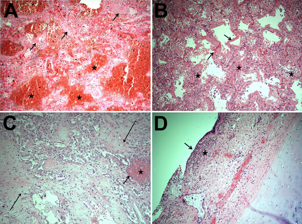
Figure 2. Results of autopsies for patients who died from influenza A(H1N1) virus infection during the 2013–14 influenza season in Florida Department of Health Region 3, Florida, USA. A) Marked intraalveolar hemorrhage (stars) and hyaline membranes (arrows) were seen during the early phase of diffuse alveolar damage and were the most common findings in the 5 autopsy cases reviewed. B) Hyaline membranes, a proteinaceous exudate replacing the alveolar walls (arrows), are prominent in this lung section, as is interstitial edema. Also, a lymphocytic infiltrating inflammation, characteristic of viral pneumonia, is shown (stars). These histologic findings correspond clinically to changes that occur during acute respiratory distress syndrome, and they occur 3–7 days after lung injury. C) The alveolar parenchyma has been replaced predominately by spindled, proliferative fibroblasts (long arrows) and hyperplastic type II pneumocytes (short arrow), indicative of the organizing phase of diffuse alveolar damage seen ≈1 week or more after lung injury. A small focus of intraalveolar hemorrhage is also present (star). D) Necrotizing tracheitis, characterized by desquamation of the tracheal columnar epithelium (arrow) and submucosal acute inflammation (star), is shown. Original magnification ×100 for all panels.