Volume 22, Number 1—January 2016
Letter
Ebola Virus Disease Complicated by Late-Onset Encephalitis and Polyarthritis, Sierra Leone
Patrick Howlett , Colin Brown, Trina Helderman, Tim Brooks, Durodamil Lisk, Gibrilla Deen, Marylou Solbrig, and Marta Lado
Figure
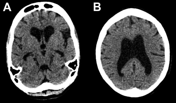
Figure. Representative axial cuts from noncontrast head computed tomography scan imaging of a 30-year-old woman with encephalitis resulting from Ebola virus infection, Sierra Leone. Images show global atrophy in keeping with nonobstructive ventriculomegaly and no periventricular low attenuation: A) subcortical atrophy; B) cortical atrophy. There was no evidence of hydrocephalus, previous stroke, or intracranial hemorrhage. A cavum septum pellucidum was noted in other images.
Page created: December 18, 2015
Page updated: December 18, 2015
Page reviewed: December 18, 2015
The conclusions, findings, and opinions expressed by authors contributing to this journal do not necessarily reflect the official position of the U.S. Department of Health and Human Services, the Public Health Service, the Centers for Disease Control and Prevention, or the authors' affiliated institutions. Use of trade names is for identification only and does not imply endorsement by any of the groups named above.