Synopses
The global epidemiology of Haemophilus ducreyi infections is poorly documented because of difficulties in confirming microbiological diagnoses. We evaluated published data on the proportion of genital and nongenital skin ulcers caused by H. ducreyi before and after introduction of syndromic management for genital ulcer disease (GUD). Before 2000, the proportion of GUD caused by H. ducreyi ranged from 0.0% to 69.0% (35 studies in 25 countries). After 2000, the proportion ranged from 0.0% to 15.0% (14 studies in 13 countries). In contrast, H. ducreyi has been recently identified as a causative agent of skin ulcers in children in the tropical regions; proportions ranged from 9.0% to 60.0% (6 studies in 4 countries). We conclude that, although there has been a sustained reduction in the proportion of GUD caused by H. ducreyi, this bacterium is increasingly recognized as a major cause of nongenital cutaneous ulcers.
| EID | González-Beiras C, Marks M, Chen CY, Roberts S, Mitjà O. Epidemiology of Haemophilus ducreyi Infections. Emerg Infect Dis. 2016;22(1):1-8. https://doi.org/10.3201/eid2201.150425 |
|---|---|
| AMA | González-Beiras C, Marks M, Chen CY, et al. Epidemiology of Haemophilus ducreyi Infections. Emerging Infectious Diseases. 2016;22(1):1-8. doi:10.3201/eid2201.150425. |
| APA | González-Beiras, C., Marks, M., Chen, C. Y., Roberts, S., & Mitjà, O. (2016). Epidemiology of Haemophilus ducreyi Infections. Emerging Infectious Diseases, 22(1), 1-8. https://doi.org/10.3201/eid2201.150425. |
Research
Multiorgan WU Polyomavirus Infection in Bone Marrow Transplant Recipient [PDF - 617 KB - 8 pages]
WU polyomavirus (WUPyV) was detected in a bone marrow transplant recipient with severe acute respiratory distress syndrome who died in 2001. Crystalline lattices of polyomavirus-like particles were observed in the patient’s lung by electron microscopy. WUPyV was detected in the lung and other tissues by real-time quantitative PCR and identified in the lung and trachea by immunohistochemistry. A subset of WUPyV-positive cells in the lung had morphologic features of macrophages. Although the role of WUPyV as a human pathogen remains unclear, these results clearly demonstrate evidence for infection of respiratory tract tissues in this patient.
| EID | Siebrasse EA, Nguyen NL, Willby MJ, Erdman DD, Menegus M, Wang D. Multiorgan WU Polyomavirus Infection in Bone Marrow Transplant Recipient. Emerg Infect Dis. 2016;22(1):24-31. https://doi.org/10.3201/eid2201.151384 |
|---|---|
| AMA | Siebrasse EA, Nguyen NL, Willby MJ, et al. Multiorgan WU Polyomavirus Infection in Bone Marrow Transplant Recipient. Emerging Infectious Diseases. 2016;22(1):24-31. doi:10.3201/eid2201.151384. |
| APA | Siebrasse, E. A., Nguyen, N. L., Willby, M. J., Erdman, D. D., Menegus, M., & Wang, D. (2016). Multiorgan WU Polyomavirus Infection in Bone Marrow Transplant Recipient. Emerging Infectious Diseases, 22(1), 24-31. https://doi.org/10.3201/eid2201.151384. |
Human Papillomavirus Prevalence and Herd Immunity after Introduction of Vaccination Program, Scotland, 2009–2013 [PDF - 527 KB - 9 pages]
In 2008, a national human papillomavirus (HPV) immunization program using a bivalent vaccine against HPV types 16 and 18 was implemented in Scotland along with a national surveillance program designed to determine the longitudinal effects of vaccination on HPV infection at the population level. Each year during 2009–2013, the surveillance program conducted HPV testing on a proportion of liquid-based cytology samples from women undergoing their first cervical screening test for precancerous cervical disease. By linking vaccination, cervical screening, and HPV testing data, over the study period we found a decline in HPV types 16 and 18, significant decreases in HPV types 31, 33, and 45 (suggesting cross-protection), and a nonsignificant increase in HPV 51. In addition, among nonvaccinated women, HPV types 16 and 18 infections were significantly lower in 2013 than in 2009. Our results preliminarily indicate herd immunity and sustained effectiveness of the bivalent vaccine on virologic outcomes at the population level.
| EID | Cameron RL, Kavanagh K, Pan J, Love J, Cuschieri K, Robertson C, et al. Human Papillomavirus Prevalence and Herd Immunity after Introduction of Vaccination Program, Scotland, 2009–2013. Emerg Infect Dis. 2016;22(1):56-64. https://doi.org/10.3201/eid2201.150736 |
|---|---|
| AMA | Cameron RL, Kavanagh K, Pan J, et al. Human Papillomavirus Prevalence and Herd Immunity after Introduction of Vaccination Program, Scotland, 2009–2013. Emerging Infectious Diseases. 2016;22(1):56-64. doi:10.3201/eid2201.150736. |
| APA | Cameron, R. L., Kavanagh, K., Pan, J., Love, J., Cuschieri, K., Robertson, C....Pollock, K. (2016). Human Papillomavirus Prevalence and Herd Immunity after Introduction of Vaccination Program, Scotland, 2009–2013. Emerging Infectious Diseases, 22(1), 56-64. https://doi.org/10.3201/eid2201.150736. |
Deaths from Plasmodium knowlesi malaria have been linked to delayed parenteral treatment. In Malaysia, early intravenous artesunate is now recommended for all severe malaria cases. We describe P. knowlesi fatalities in Sabah, Malaysia, during 2012–2014 and report species-specific fatality rates based on 2010–2014 case notifications. Sixteen malaria-associated deaths (caused by PCR-confirmed P. knowlesi [7], P. falciparum [7], and P. vivax [1] and microscopy-diagnosed “P. malariae” [1]) were reported during 2012–2014. Six patients with severe P. knowlesi malaria received intravenous artesunate at hospital admission. For persons >15 years of age, overall fatality rates during 2010–2014 were 3.4, 4.2, and 1.0 deaths/1,000 P. knowlesi, P. falciparum, and P. vivax notifications, respectively; P. knowlesi–associated fatality rates fell from 9.2 to1.6 deaths/1,000 notifications. No P. knowlesi–associated deaths occurred among children, despite 373 notified cases. Although P. knowlesi malaria incidence is rising, the notification-fatality rate has decreased, likely due to improved use of intravenous artesunate.
| EID | Rajahram GS, Barber BE, William T, Grigg MJ, Menon J, Yeo TW, et al. Falling Plasmodium knowlesi Malaria Death Rate among Adults despite Rising Incidence, Sabah, Malaysia, 2010–2014. Emerg Infect Dis. 2016;22(1):41-48. https://doi.org/10.3201/eid2201.151305 |
|---|---|
| AMA | Rajahram GS, Barber BE, William T, et al. Falling Plasmodium knowlesi Malaria Death Rate among Adults despite Rising Incidence, Sabah, Malaysia, 2010–2014. Emerging Infectious Diseases. 2016;22(1):41-48. doi:10.3201/eid2201.151305. |
| APA | Rajahram, G. S., Barber, B. E., William, T., Grigg, M. J., Menon, J., Yeo, T. W....Anstey, N. M. (2016). Falling Plasmodium knowlesi Malaria Death Rate among Adults despite Rising Incidence, Sabah, Malaysia, 2010–2014. Emerging Infectious Diseases, 22(1), 41-48. https://doi.org/10.3201/eid2201.151305. |
Multifacility Outbreak of Middle East Respiratory Syndrome in Taif, Saudi Arabia [PDF - 1.12 MB - 9 pages]
Middle East respiratory syndrome (MERS) coronavirus (MERS-CoV) is a novel respiratory pathogen first reported in 2012. During September 2014–January 2015, an outbreak of 38 cases of MERS was reported from 4 healthcare facilities in Taif, Saudi Arabia; 21 of the 38 case-patients died. Clinical and public health records showed that 13 patients were healthcare personnel (HCP). Fifteen patients, including 4 HCP, were associated with 1 dialysis unit. Three additional HCP in this dialysis unit had serologic evidence of MERS-CoV infection. Viral RNA was amplified from acute-phase serum specimens of 15 patients, and full spike gene-coding sequencing was obtained from 10 patients who formed a discrete cluster; sequences from specimens of 9 patients were closely related. Similar gene sequences among patients unlinked by time or location suggest unrecognized viral transmission. Circulation persisted in multiple healthcare settings over an extended period, underscoring the importance of strengthening MERS-CoV surveillance and infection-control practices.
| EID | Assiri AM, Abedi GR, Saeed AA, Abdalla MA, al-Masry M, Choudhry A, et al. Multifacility Outbreak of Middle East Respiratory Syndrome in Taif, Saudi Arabia. Emerg Infect Dis. 2016;22(1):32-40. https://doi.org/10.3201/eid2201.151370 |
|---|---|
| AMA | Assiri AM, Abedi GR, Saeed AA, et al. Multifacility Outbreak of Middle East Respiratory Syndrome in Taif, Saudi Arabia. Emerging Infectious Diseases. 2016;22(1):32-40. doi:10.3201/eid2201.151370. |
| APA | Assiri, A. M., Abedi, G. R., Saeed, A. A., Abdalla, M. A., al-Masry, M., Choudhry, A....Watson, J. T. (2016). Multifacility Outbreak of Middle East Respiratory Syndrome in Taif, Saudi Arabia. Emerging Infectious Diseases, 22(1), 32-40. https://doi.org/10.3201/eid2201.151370. |
Risk Factors for Primary Middle East Respiratory Syndrome Coronavirus Illness in Humans, Saudi Arabia, 2014 [PDF - 449 KB - 7 pages]
Risk factors for primary Middle East respiratory syndrome coronavirus (MERS-CoV) illness in humans are incompletely understood. We identified all primary MERS-CoV cases reported in Saudi Arabia during March–November 2014 by excluding those with history of exposure to other cases of MERS-CoV or acute respiratory illness of unknown cause or exposure to healthcare settings within 14 days before illness onset. Using a case–control design, we assessed differences in underlying medical conditions and environmental exposures among primary case-patients and 2–4 controls matched by age, sex, and neighborhood. Using multivariable analysis, we found that direct exposure to dromedary camels during the 2 weeks before illness onset, as well as diabetes mellitus, heart disease, and smoking, were each independently associated with MERS-CoV illness. Further investigation is needed to better understand animal-to-human transmission of MERS-CoV.
| EID | Alraddadi BM, Watson JT, Almarashi A, Abedi GR, Turkistani A, Sadran M, et al. Risk Factors for Primary Middle East Respiratory Syndrome Coronavirus Illness in Humans, Saudi Arabia, 2014. Emerg Infect Dis. 2016;22(1):49-55. https://doi.org/10.3201/eid2201.151340 |
|---|---|
| AMA | Alraddadi BM, Watson JT, Almarashi A, et al. Risk Factors for Primary Middle East Respiratory Syndrome Coronavirus Illness in Humans, Saudi Arabia, 2014. Emerging Infectious Diseases. 2016;22(1):49-55. doi:10.3201/eid2201.151340. |
| APA | Alraddadi, B. M., Watson, J. T., Almarashi, A., Abedi, G. R., Turkistani, A., Sadran, M....Madani, T. A. (2016). Risk Factors for Primary Middle East Respiratory Syndrome Coronavirus Illness in Humans, Saudi Arabia, 2014. Emerging Infectious Diseases, 22(1), 49-55. https://doi.org/10.3201/eid2201.151340. |
Waterborne Elizabethkingia meningoseptica in Adult Critical Care [PDF - 594 KB - 9 pages]
Elizabethkingia meningoseptica is an infrequent colonizer of the respiratory tract; its pathogenicity is uncertain. In the context of a 22-month outbreak of E. meningoseptica acquisition affecting 30 patients in a London, UK, critical care unit (3% attack rate) we derived a measure of attributable morbidity and determined whether E. meningoseptica is an emerging nosocomial pathogen. We found monomicrobial E. meningoseptica acquisition (n = 13) to have an attributable morbidity rate of 54% (systemic inflammatory response syndrome >2, rising C-reactive protein, new radiographic changes), suggesting that E. meningoseptica is a pathogen. Epidemiologic and molecular evidence showed acquisition was water-source–associated in critical care but identified numerous other E. meningoseptica strains, indicating more widespread distribution than previously considered. Analysis of changes in gram-negative speciation rates across a wider London hospital network suggests this outbreak, and possibly other recently reported outbreaks, might reflect improved diagnostics and that E. meningoseptica thus is a pseudo-emerging pathogen.
| EID | Moore L, Owens DS, Jepson A, Turton JF, Ashworth S, Donaldson H, et al. Waterborne Elizabethkingia meningoseptica in Adult Critical Care. Emerg Infect Dis. 2016;22(1):9-17. https://doi.org/10.3201/eid2201.150139 |
|---|---|
| AMA | Moore L, Owens DS, Jepson A, et al. Waterborne Elizabethkingia meningoseptica in Adult Critical Care. Emerging Infectious Diseases. 2016;22(1):9-17. doi:10.3201/eid2201.150139. |
| APA | Moore, L., Owens, D. S., Jepson, A., Turton, J. F., Ashworth, S., Donaldson, H....Holmes, A. H. (2016). Waterborne Elizabethkingia meningoseptica in Adult Critical Care. Emerging Infectious Diseases, 22(1), 9-17. https://doi.org/10.3201/eid2201.150139. |
Human Papillomavirus Vaccination at a Time of Changing Sexual Behavior [PDF - 390 KB - 6 pages]
Human papillomavirus (HPV) prevalence varies widely worldwide. We used a transmission model to show links between age-specific sexual patterns and HPV vaccination effectiveness. We considered rural India and the United States as examples of 2 heterosexual populations with traditional age-specific sexual behavior and gender-similar age-specific sexual behavior, respectively. We simulated these populations by using age-specific rates of sexual activity and age differences between sexual partners and found that transitions from traditional to gender-similar sexual behavior in women <35 years of age can result in increased (2.6-fold in our study) HPV16 prevalence. Our model shows that reductions in HPV16 prevalence are larger if vaccination occurs in populations before transitions in sexual behavior and that increased risk for HPV infection attributable to transition is preventable by early vaccination. Our study highlights the importance of using time-limited opportunities to introduce HPV vaccination in traditional populations before changes in age-specific sexual patterns occur.
| EID | Baussano I, Lazzarato F, Brisson M, Franceschi S. Human Papillomavirus Vaccination at a Time of Changing Sexual Behavior. Emerg Infect Dis. 2016;22(1):18-23. https://doi.org/10.3201/eid2201.150791 |
|---|---|
| AMA | Baussano I, Lazzarato F, Brisson M, et al. Human Papillomavirus Vaccination at a Time of Changing Sexual Behavior. Emerging Infectious Diseases. 2016;22(1):18-23. doi:10.3201/eid2201.150791. |
| APA | Baussano, I., Lazzarato, F., Brisson, M., & Franceschi, S. (2016). Human Papillomavirus Vaccination at a Time of Changing Sexual Behavior. Emerging Infectious Diseases, 22(1), 18-23. https://doi.org/10.3201/eid2201.150791. |
Dispatches
Severe Community-Acquired Bloodstream Infection with Acinetobacter ursingii in Person who Injects Drugs [PDF - 374 KB - 4 pages]
We report a community-acquired bloodstream infection with Acinteobacter ursingii in an HIV-negative woman who injected drugs. The infection was successfully treated with meropenem. Species identification was performed by using matrix-assisted laser desorption/ionization time-of-flight mass spectrometry. Improved identification of Acinetobacter spp. by using this method will help identify clinical effects of this underdiagnosed pathogen.
| EID | Salzer H, Rolling T, Schmiedel S, Klupp E, Lange C, Seifert H. Severe Community-Acquired Bloodstream Infection with Acinetobacter ursingii in Person who Injects Drugs. Emerg Infect Dis. 2016;22(1):134-137. https://doi.org/10.3201/eid2201.151298 |
|---|---|
| AMA | Salzer H, Rolling T, Schmiedel S, et al. Severe Community-Acquired Bloodstream Infection with Acinetobacter ursingii in Person who Injects Drugs. Emerging Infectious Diseases. 2016;22(1):134-137. doi:10.3201/eid2201.151298. |
| APA | Salzer, H., Rolling, T., Schmiedel, S., Klupp, E., Lange, C., & Seifert, H. (2016). Severe Community-Acquired Bloodstream Infection with Acinetobacter ursingii in Person who Injects Drugs. Emerging Infectious Diseases, 22(1), 134-137. https://doi.org/10.3201/eid2201.151298. |
Avian Influenza A(H7N9) Virus Infection in 2 Travelers Returning from China to Canada, January 2015 [PDF - 483 KB - 4 pages]
In January 2015, British Columbia, Canada, reported avian influenza A(H7N9) virus infection in 2 travelers returning from China who sought outpatient care for typical influenza-like illness. There was no further spread, but serosurvey findings showed broad population susceptibility to H7N9 virus. Travel history and timely notification are critical to emerging pathogen detection and response.
| EID | Skowronski D, Chambers C, Gustafson R, Purych DB, Tang P, Bastien N, et al. Avian Influenza A(H7N9) Virus Infection in 2 Travelers Returning from China to Canada, January 2015. Emerg Infect Dis. 2016;22(1):71-74. https://doi.org/10.3201/eid2201.151330 |
|---|---|
| AMA | Skowronski D, Chambers C, Gustafson R, et al. Avian Influenza A(H7N9) Virus Infection in 2 Travelers Returning from China to Canada, January 2015. Emerging Infectious Diseases. 2016;22(1):71-74. doi:10.3201/eid2201.151330. |
| APA | Skowronski, D., Chambers, C., Gustafson, R., Purych, D. B., Tang, P., Bastien, N....Li, Y. (2016). Avian Influenza A(H7N9) Virus Infection in 2 Travelers Returning from China to Canada, January 2015. Emerging Infectious Diseases, 22(1), 71-74. https://doi.org/10.3201/eid2201.151330. |
Variations in Spike Glycoprotein Gene of MERS-CoV, South Korea, 2015 [PDF - 643 KB - 5 pages]
An outbreak of nosocomial infections with Middle East respiratory syndrome coronavirus occurred in South Korea in May 2015. Spike glycoprotein genes of virus strains from South Korea were closely related to those of strains from Riyadh, Saudi Arabia. However, virus strains from South Korea showed strain-specific variations.
| EID | Kim D, Kim Y, Park S, Yun M, Yang J, Kang H, et al. Variations in Spike Glycoprotein Gene of MERS-CoV, South Korea, 2015. Emerg Infect Dis. 2016;22(1):100-104. https://doi.org/10.3201/eid2201.151055 |
|---|---|
| AMA | Kim D, Kim Y, Park S, et al. Variations in Spike Glycoprotein Gene of MERS-CoV, South Korea, 2015. Emerging Infectious Diseases. 2016;22(1):100-104. doi:10.3201/eid2201.151055. |
| APA | Kim, D., Kim, Y., Park, S., Yun, M., Yang, J., Kang, H....Kim, S. (2016). Variations in Spike Glycoprotein Gene of MERS-CoV, South Korea, 2015. Emerging Infectious Diseases, 22(1), 100-104. https://doi.org/10.3201/eid2201.151055. |
Asymptomatic Lymphogranuloma Venereum in Men who Have Sex with Men, United Kingdom [PDF - 398 KB - 5 pages]
We investigated prevalence of lymphogranuloma venereum (LGV) among men who have sex with men who were tested for chlamydia at 12 clinics in the United Kingdom during 10 weeks in 2012. Of 713 men positive for Chlamydia trachomatis, 66 (9%) had LGV serovars; 15 (27%) of 55 for whom data were available were asymptomatic.
| EID | Saxon C, Hughes G, Ison C. Asymptomatic Lymphogranuloma Venereum in Men who Have Sex with Men, United Kingdom. Emerg Infect Dis. 2016;22(1):112-116. https://doi.org/10.3201/eid2201.141867 |
|---|---|
| AMA | Saxon C, Hughes G, Ison C. Asymptomatic Lymphogranuloma Venereum in Men who Have Sex with Men, United Kingdom. Emerging Infectious Diseases. 2016;22(1):112-116. doi:10.3201/eid2201.141867. |
| APA | Saxon, C., Hughes, G., & Ison, C. (2016). Asymptomatic Lymphogranuloma Venereum in Men who Have Sex with Men, United Kingdom. Emerging Infectious Diseases, 22(1), 112-116. https://doi.org/10.3201/eid2201.141867. |
Rift Valley Fever Virus among Wild Ruminants, Etosha National Park, Namibia, 2011 [PDF - 304 KB - 3 pages]
After a May 2011 outbreak of Rift Valley fever among livestock northeast of Etosha National Park, Namibia, wild ruminants in the park were tested for the virus. Antibodies were detected in springbok, wildebeest, and black-faced impala, and viral RNA was detected in springbok. Seroprevalence was high, and immune response was long lasting.
| EID | Dondona A, Aschenborn O, Pinoni C, Di Gialleonardo L, Maseke A, Bortone G, et al. Rift Valley Fever Virus among Wild Ruminants, Etosha National Park, Namibia, 2011. Emerg Infect Dis. 2016;22(1):128-130. https://doi.org/10.3201/eid2201.150725 |
|---|---|
| AMA | Dondona A, Aschenborn O, Pinoni C, et al. Rift Valley Fever Virus among Wild Ruminants, Etosha National Park, Namibia, 2011. Emerging Infectious Diseases. 2016;22(1):128-130. doi:10.3201/eid2201.150725. |
| APA | Dondona, A., Aschenborn, O., Pinoni, C., Di Gialleonardo, L., Maseke, A., Bortone, G....Monaco, F. (2016). Rift Valley Fever Virus among Wild Ruminants, Etosha National Park, Namibia, 2011. Emerging Infectious Diseases, 22(1), 128-130. https://doi.org/10.3201/eid2201.150725. |
Identification of Source of Brucella suis Infection in Human by Whole-Genome Sequencing, United States and Tonga [PDF - 519 KB - 4 pages]
Brucella suis infection was diagnosed in a man from Tonga, Polynesia, who had butchered swine in Oregon, USA. Although the US commercial swine herd is designated brucellosis-free, exposure history suggested infection from commercial pigs. We used whole-genome sequencing to determine that the man was infected in Tonga, averting a field investigation.
| EID | Quance C, Robbe-Austerman S, Stuber T, Brignole T, DeBess EE, Boyd L, et al. Identification of Source of Brucella suis Infection in Human by Whole-Genome Sequencing, United States and Tonga. Emerg Infect Dis. 2016;22(1):79-82. https://doi.org/10.3201/eid2201.150843 |
|---|---|
| AMA | Quance C, Robbe-Austerman S, Stuber T, et al. Identification of Source of Brucella suis Infection in Human by Whole-Genome Sequencing, United States and Tonga. Emerging Infectious Diseases. 2016;22(1):79-82. doi:10.3201/eid2201.150843. |
| APA | Quance, C., Robbe-Austerman, S., Stuber, T., Brignole, T., DeBess, E. E., Boyd, L....Erdman, M. M. (2016). Identification of Source of Brucella suis Infection in Human by Whole-Genome Sequencing, United States and Tonga. Emerging Infectious Diseases, 22(1), 79-82. https://doi.org/10.3201/eid2201.150843. |
Seroepidemiology of Human Enterovirus 71 Infection among Children, Cambodia [PDF - 386 KB - 4 pages]
Enterovirus 71 is reported to have emerged in Cambodia in 2012; at least 54 children with severe encephalitis died during that outbreak. We used serum samples collected during 2000–2011 to show that the virus had been widespread in the country for at least a decade before the 2012 outbreak.
| EID | Horwood PF, Andronico A, Tarantola A, Salje H, Duong V, Mey C, et al. Seroepidemiology of Human Enterovirus 71 Infection among Children, Cambodia. Emerg Infect Dis. 2016;22(1):92-95. https://doi.org/10.3201/eid2201.151323 |
|---|---|
| AMA | Horwood PF, Andronico A, Tarantola A, et al. Seroepidemiology of Human Enterovirus 71 Infection among Children, Cambodia. Emerging Infectious Diseases. 2016;22(1):92-95. doi:10.3201/eid2201.151323. |
| APA | Horwood, P. F., Andronico, A., Tarantola, A., Salje, H., Duong, V., Mey, C....Buchy, P. (2016). Seroepidemiology of Human Enterovirus 71 Infection among Children, Cambodia. Emerging Infectious Diseases, 22(1), 92-95. https://doi.org/10.3201/eid2201.151323. |
Effectiveness of Ring Vaccination as Control Strategy for Ebola Virus Disease [PDF - 463 KB - 4 pages]
Using an Ebola virus disease transmission model, we found that addition of ring vaccination at the outset of the West Africa epidemic might not have led to containment of this disease. However, in later stages of the epidemic or in outbreaks with less intense transmission or more effective control, this strategy could help eliminate the disease.
| EID | Kucharski AJ, Eggo RM, Watson C, Camacho A, Funk S, Edmunds W. Effectiveness of Ring Vaccination as Control Strategy for Ebola Virus Disease. Emerg Infect Dis. 2016;22(1):105-108. https://doi.org/10.3201/eid2201.151410 |
|---|---|
| AMA | Kucharski AJ, Eggo RM, Watson C, et al. Effectiveness of Ring Vaccination as Control Strategy for Ebola Virus Disease. Emerging Infectious Diseases. 2016;22(1):105-108. doi:10.3201/eid2201.151410. |
| APA | Kucharski, A. J., Eggo, R. M., Watson, C., Camacho, A., Funk, S., & Edmunds, W. (2016). Effectiveness of Ring Vaccination as Control Strategy for Ebola Virus Disease. Emerging Infectious Diseases, 22(1), 105-108. https://doi.org/10.3201/eid2201.151410. |
Increase in Sexually Transmitted Infections among Men Who Have Sex with Men, England, 2014 [PDF - 548 KB - 4 pages]
Surveillance data from sexual health clinics indicate recent increases in sexually transmitted infections, particularly among men who have sex with men. The largest annual increase in syphilis diagnoses in a decade was reported in 2014. Less condom use may be the primary reason for these increases.
| EID | Mohammed H, Mitchell H, Sile B, Duffell S, Nardone A, Hughes G. Increase in Sexually Transmitted Infections among Men Who Have Sex with Men, England, 2014. Emerg Infect Dis. 2016;22(1):88-91. https://doi.org/10.3201/eid2201.151331 |
|---|---|
| AMA | Mohammed H, Mitchell H, Sile B, et al. Increase in Sexually Transmitted Infections among Men Who Have Sex with Men, England, 2014. Emerging Infectious Diseases. 2016;22(1):88-91. doi:10.3201/eid2201.151331. |
| APA | Mohammed, H., Mitchell, H., Sile, B., Duffell, S., Nardone, A., & Hughes, G. (2016). Increase in Sexually Transmitted Infections among Men Who Have Sex with Men, England, 2014. Emerging Infectious Diseases, 22(1), 88-91. https://doi.org/10.3201/eid2201.151331. |
Rapid Emergence and Clonal Dissemination of CTX-M-15–Producing Salmonella enterica Serotype Virchow, South Korea [PDF - 310 KB - 3 pages]
The prevalence of cefotaxime-resistant Salmonella enterica serotype Virchow has dramatically increased in South Korea since the first isolation in 2011. Of 68 isolates collected over 10 years, 28 cefotaxime-resistant isolates harbored the blaCTX-M-15 extended-spectrum β-lactamase gene and were closely related genetically, demonstrating the clonal dissemination of CTX-M-15–producing Salmonella Virchow in South Korea.
| EID | Kim J, Yun Y, Kim S, Jeon S, Lee D, Chung G, et al. Rapid Emergence and Clonal Dissemination of CTX-M-15–Producing Salmonella enterica Serotype Virchow, South Korea. Emerg Infect Dis. 2016;22(1):68-70. https://doi.org/10.3201/eid2201.151220 |
|---|---|
| AMA | Kim J, Yun Y, Kim S, et al. Rapid Emergence and Clonal Dissemination of CTX-M-15–Producing Salmonella enterica Serotype Virchow, South Korea. Emerging Infectious Diseases. 2016;22(1):68-70. doi:10.3201/eid2201.151220. |
| APA | Kim, J., Yun, Y., Kim, S., Jeon, S., Lee, D., Chung, G....Kim, J. (2016). Rapid Emergence and Clonal Dissemination of CTX-M-15–Producing Salmonella enterica Serotype Virchow, South Korea. Emerging Infectious Diseases, 22(1), 68-70. https://doi.org/10.3201/eid2201.151220. |
Increased Risk for ESBL-Producing Bacteria from Co-administration of Loperamide and Antimicrobial Drugs for Travelers’ Diarrhea [PDF - 465 KB - 4 pages]
Antimicrobial drug treatment of travelers’ diarrhea is known to increase the risk for colonization with extended-spectrum β-lactamase-producing Enterobacteriaceae. Among 288 travelers with travelers’ diarrhea, the colonization rate without medications was 21%. For treatment with loperamide only, the rate was 20%; with antimicrobial drugs alone, 40%; and with loperamide and antimicrobial drugs, 71%.
| EID | Kantele A, Mero S, Kirveskari J, Lääveri T. Increased Risk for ESBL-Producing Bacteria from Co-administration of Loperamide and Antimicrobial Drugs for Travelers’ Diarrhea. Emerg Infect Dis. 2016;22(1):117-120. https://doi.org/10.3201/eid2201.151272 |
|---|---|
| AMA | Kantele A, Mero S, Kirveskari J, et al. Increased Risk for ESBL-Producing Bacteria from Co-administration of Loperamide and Antimicrobial Drugs for Travelers’ Diarrhea. Emerging Infectious Diseases. 2016;22(1):117-120. doi:10.3201/eid2201.151272. |
| APA | Kantele, A., Mero, S., Kirveskari, J., & Lääveri, T. (2016). Increased Risk for ESBL-Producing Bacteria from Co-administration of Loperamide and Antimicrobial Drugs for Travelers’ Diarrhea. Emerging Infectious Diseases, 22(1), 117-120. https://doi.org/10.3201/eid2201.151272. |
Autochthonous Nocardia cerradoensis Infection in Humans, Spain, 2011 and 2014 [PDF - 346 KB - 3 pages]
Nocardia cerradoensis was first isolated in 2003 in the El Cerrado region of Brazil; since then, only 2 human infections, in France and Spain, have been reported. We describe 3 autochthonous cases in residents of Spain during 2011 and 2014. Together these cases support the idea of an emerging global pathogenic microorganism.
| EID | Ercibengoa M, Pérez-Trallero E, Marimón J. Autochthonous Nocardia cerradoensis Infection in Humans, Spain, 2011 and 2014. Emerg Infect Dis. 2016;22(1):109-111. https://doi.org/10.3201/eid2201.150771 |
|---|---|
| AMA | Ercibengoa M, Pérez-Trallero E, Marimón J. Autochthonous Nocardia cerradoensis Infection in Humans, Spain, 2011 and 2014. Emerging Infectious Diseases. 2016;22(1):109-111. doi:10.3201/eid2201.150771. |
| APA | Ercibengoa, M., Pérez-Trallero, E., & Marimón, J. (2016). Autochthonous Nocardia cerradoensis Infection in Humans, Spain, 2011 and 2014. Emerging Infectious Diseases, 22(1), 109-111. https://doi.org/10.3201/eid2201.150771. |
Hemagglutinin Gene Clade 3C.2a Influenza A(H3N2) Viruses, Alachua County, Florida, USA, 2014–15 [PDF - 316 KB - 3 pages]
Influenza A(H3N2) strains isolated during 2014–15 in Alachua County, Florida, USA, belonged to hemagglutinin gene clade 3C.2a. High rates of influenza-like illness and confirmed influenza cases in children were associated with a decrease in estimated vaccine effectiveness. Illnesses were milder than in 2013–14; severe cases were concentrated in elderly patients with underlying diseases.
| EID | Lednicky J, Iovine NM, Brew J, Loeb J, Sugimoto JD, Rand KH, et al. Hemagglutinin Gene Clade 3C.2a Influenza A(H3N2) Viruses, Alachua County, Florida, USA, 2014–15. Emerg Infect Dis. 2016;22(1):121-123. https://doi.org/10.3201/eid2201.151019 |
|---|---|
| AMA | Lednicky J, Iovine NM, Brew J, et al. Hemagglutinin Gene Clade 3C.2a Influenza A(H3N2) Viruses, Alachua County, Florida, USA, 2014–15. Emerging Infectious Diseases. 2016;22(1):121-123. doi:10.3201/eid2201.151019. |
| APA | Lednicky, J., Iovine, N. M., Brew, J., Loeb, J., Sugimoto, J. D., Rand, K. H....Morris, J. (2016). Hemagglutinin Gene Clade 3C.2a Influenza A(H3N2) Viruses, Alachua County, Florida, USA, 2014–15. Emerging Infectious Diseases, 22(1), 121-123. https://doi.org/10.3201/eid2201.151019. |
Factors Related to Fetal Death in Pregnant Women with Cholera, Haiti, 2011–2014 [PDF - 348 KB - 4 pages]
We assessed risk factors for fetal death during cholera infection and effect of treatment changes on these deaths. Third trimester gestation, younger maternal age, severe dehydration, and vomiting were risk factors. Changes in treatment had limited effects on fetal death, highlighting the need for prevention and evidence-based treatment.
| EID | Schillberg E, Ariti C, Bryson L, Delva-Senat R, Price D, GrandPierre R, et al. Factors Related to Fetal Death in Pregnant Women with Cholera, Haiti, 2011–2014. Emerg Infect Dis. 2016;22(1):124-127. https://doi.org/10.3201/eid2201.151078 |
|---|---|
| AMA | Schillberg E, Ariti C, Bryson L, et al. Factors Related to Fetal Death in Pregnant Women with Cholera, Haiti, 2011–2014. Emerging Infectious Diseases. 2016;22(1):124-127. doi:10.3201/eid2201.151078. |
| APA | Schillberg, E., Ariti, C., Bryson, L., Delva-Senat, R., Price, D., GrandPierre, R....Lenglet, A. (2016). Factors Related to Fetal Death in Pregnant Women with Cholera, Haiti, 2011–2014. Emerging Infectious Diseases, 22(1), 124-127. https://doi.org/10.3201/eid2201.151078. |
Surveillance of Bacterial Meningitis, Ethiopia, 2012–2013 [PDF - 514 KB - 4 pages]
Among 139 patients with suspected bacterial meningitis in Ethiopia, 2012–2013, meningococci (19.4%) and pneumococci (12.9%) were the major disease-causing organisms. Meningococcal serogroups detected were A (n = 11), W (n = 7), C (n = 1), and X (n = 1). Affordable, multivalent meningitis vaccines for the African meningitis belt are urgently needed.
| EID | Mihret W, Lema T, Merid Y, Kassu A, Abebe W, Moges B, et al. Surveillance of Bacterial Meningitis, Ethiopia, 2012–2013. Emerg Infect Dis. 2016;22(1):75-78. https://doi.org/10.3201/eid2201.150432 |
|---|---|
| AMA | Mihret W, Lema T, Merid Y, et al. Surveillance of Bacterial Meningitis, Ethiopia, 2012–2013. Emerging Infectious Diseases. 2016;22(1):75-78. doi:10.3201/eid2201.150432. |
| APA | Mihret, W., Lema, T., Merid, Y., Kassu, A., Abebe, W., Moges, B....Norheim, G. (2016). Surveillance of Bacterial Meningitis, Ethiopia, 2012–2013. Emerging Infectious Diseases, 22(1), 75-78. https://doi.org/10.3201/eid2201.150432. |
Porcine Epidemic Diarrhea Virus and Discovery of a Recombinant Swine Enteric Coronavirus, Italy [PDF - 610 KB - 5 pages]
Porcine epidemic diarrhea virus (PEDV) has been detected sporadically in Italy since the 1990s. We report the phylogenetic relationship of swine enteric coronaviruses collected in Italy during 2007–2014 and identify a drastic shift in PEDV strain variability and a new swine enteric coronavirus generated by recombination of transmissible gastroenteritis virus and PEDV.
| EID | Boniotti M, Papetti A, Lavazza A, Alborali G, Sozzi E, Chiapponi C, et al. Porcine Epidemic Diarrhea Virus and Discovery of a Recombinant Swine Enteric Coronavirus, Italy. Emerg Infect Dis. 2016;22(1):83-87. https://doi.org/10.3201/eid2201.150544 |
|---|---|
| AMA | Boniotti M, Papetti A, Lavazza A, et al. Porcine Epidemic Diarrhea Virus and Discovery of a Recombinant Swine Enteric Coronavirus, Italy. Emerging Infectious Diseases. 2016;22(1):83-87. doi:10.3201/eid2201.150544. |
| APA | Boniotti, M., Papetti, A., Lavazza, A., Alborali, G., Sozzi, E., Chiapponi, C....Marthaler, D. (2016). Porcine Epidemic Diarrhea Virus and Discovery of a Recombinant Swine Enteric Coronavirus, Italy. Emerging Infectious Diseases, 22(1), 83-87. https://doi.org/10.3201/eid2201.150544. |
Outbreak of Panton-Valentine Leukocidin–Associated Methicillin-Susceptible Staphylococcus aureus Infection in a Rugby Team, France, 2010–2011 [PDF - 412 KB - 4 pages]
Staphylococcus aureus strains that produce Panton-Valentine leukocidin are known to cause community infections. We describe an outbreak of skin abscesses caused by Panton-Valentine leukocidin–producing methicillin-susceptible S. aureus (clonal complex 121) in a professional rugby team in France during July 2010–February 2011. Eight team members were carriers; 7 had skin abscesses.
| EID | Couvé-Deacon E, Tristan A, Pestourie N, Faure C, Doffoel-Hantz V, Garnier F, et al. Outbreak of Panton-Valentine Leukocidin–Associated Methicillin-Susceptible Staphylococcus aureus Infection in a Rugby Team, France, 2010–2011. Emerg Infect Dis. 2016;22(1):96-99. https://doi.org/10.3201/eid2201.150597 |
|---|---|
| AMA | Couvé-Deacon E, Tristan A, Pestourie N, et al. Outbreak of Panton-Valentine Leukocidin–Associated Methicillin-Susceptible Staphylococcus aureus Infection in a Rugby Team, France, 2010–2011. Emerging Infectious Diseases. 2016;22(1):96-99. doi:10.3201/eid2201.150597. |
| APA | Couvé-Deacon, E., Tristan, A., Pestourie, N., Faure, C., Doffoel-Hantz, V., Garnier, F....Ploy, M. (2016). Outbreak of Panton-Valentine Leukocidin–Associated Methicillin-Susceptible Staphylococcus aureus Infection in a Rugby Team, France, 2010–2011. Emerging Infectious Diseases, 22(1), 96-99. https://doi.org/10.3201/eid2201.150597. |
Decline in Decreased Cephalosporin Susceptibility and Increase in Azithromycin Resistance in Neisseria gonorrhoeae, Canada [PDF - 377 KB - 3 pages]
Antimicrobial resistance profiles were determined for Neisseria gonorrhoeae strains isolated in Canada during 2010–2014. The proportion of isolates with decreased susceptibility to cephalosporins declined significantly between 2011 and 2014, whereas azithromycin resistance increased significantly during that period. Continued surveillance of antimicrobial drug susceptibilities is imperative to inform treatment guidelines.
| EID | Martin I, Sawatzky P, Liu G, Allen V, Lefebvre B, Hoang L, et al. Decline in Decreased Cephalosporin Susceptibility and Increase in Azithromycin Resistance in Neisseria gonorrhoeae, Canada. Emerg Infect Dis. 2016;22(1):65-67. https://doi.org/10.3201/eid2201.151247 |
|---|---|
| AMA | Martin I, Sawatzky P, Liu G, et al. Decline in Decreased Cephalosporin Susceptibility and Increase in Azithromycin Resistance in Neisseria gonorrhoeae, Canada. Emerging Infectious Diseases. 2016;22(1):65-67. doi:10.3201/eid2201.151247. |
| APA | Martin, I., Sawatzky, P., Liu, G., Allen, V., Lefebvre, B., Hoang, L....Mulvey, M. (2016). Decline in Decreased Cephalosporin Susceptibility and Increase in Azithromycin Resistance in Neisseria gonorrhoeae, Canada. Emerging Infectious Diseases, 22(1), 65-67. https://doi.org/10.3201/eid2201.151247. |
Legionnaires’ Disease in South Africa, 2012–2014 [PDF - 417 KB - 3 pages]
During June 2012–September 2014, we tested patients with severe respiratory illness for Legionella spp. infection and conducted a retrospective epidemiologic investigation. Of 1,805 patients tested, Legionella was detected in samples of 21 (1.2%); most were adults who had HIV or tuberculosis infections and were inappropriately treated for Legionella.
| EID | Wolter N, Carrim M, Tempia S, Cohen C, Walaza S, Sahr P, et al. Legionnaires’ Disease in South Africa, 2012–2014. Emerg Infect Dis. 2016;22(1):131-133. https://doi.org/10.3201/eid2201.150972 |
|---|---|
| AMA | Wolter N, Carrim M, Tempia S, et al. Legionnaires’ Disease in South Africa, 2012–2014. Emerging Infectious Diseases. 2016;22(1):131-133. doi:10.3201/eid2201.150972. |
| APA | Wolter, N., Carrim, M., Tempia, S., Cohen, C., Walaza, S., Sahr, P....von Gottberg, A. (2016). Legionnaires’ Disease in South Africa, 2012–2014. Emerging Infectious Diseases, 22(1), 131-133. https://doi.org/10.3201/eid2201.150972. |
Letters
Louseborne Relapsing Fever in Young Migrants, Sicily, Italy, July–September 2015 [PDF - 273 KB - 2 pages]
| EID | Ciervo A, Mancini F, Di Bernardo F, Giammanco A, Vitale G, Dones P, et al. Louseborne Relapsing Fever in Young Migrants, Sicily, Italy, July–September 2015. Emerg Infect Dis. 2016;22(1):152-153. https://doi.org/10.3201/eid2201.151580 |
|---|---|
| AMA | Ciervo A, Mancini F, Di Bernardo F, et al. Louseborne Relapsing Fever in Young Migrants, Sicily, Italy, July–September 2015. Emerging Infectious Diseases. 2016;22(1):152-153. doi:10.3201/eid2201.151580. |
| APA | Ciervo, A., Mancini, F., Di Bernardo, F., Giammanco, A., Vitale, G., Dones, P....Rezza, G. (2016). Louseborne Relapsing Fever in Young Migrants, Sicily, Italy, July–September 2015. Emerging Infectious Diseases, 22(1), 152-153. https://doi.org/10.3201/eid2201.151580. |
Ebola Virus Disease Complicated by Late-Onset Encephalitis and Polyarthritis, Sierra Leone [PDF - 293 KB - 3 pages]
| EID | Howlett P, Brown C, Helderman T, Brooks T, Lisk D, Deen G, et al. Ebola Virus Disease Complicated by Late-Onset Encephalitis and Polyarthritis, Sierra Leone. Emerg Infect Dis. 2016;22(1):150-152. https://doi.org/10.3201/eid2201.151212 |
|---|---|
| AMA | Howlett P, Brown C, Helderman T, et al. Ebola Virus Disease Complicated by Late-Onset Encephalitis and Polyarthritis, Sierra Leone. Emerging Infectious Diseases. 2016;22(1):150-152. doi:10.3201/eid2201.151212. |
| APA | Howlett, P., Brown, C., Helderman, T., Brooks, T., Lisk, D., Deen, G....Lado, M. (2016). Ebola Virus Disease Complicated by Late-Onset Encephalitis and Polyarthritis, Sierra Leone. Emerging Infectious Diseases, 22(1), 150-152. https://doi.org/10.3201/eid2201.151212. |
New Clinical Strain of Neisseria gonorrhoeae with Decreased Susceptibility to Ceftriaxone, Japan [PDF - 353 KB - 3 pages]
| EID | Deguchi T, Yasuda M, Hatazaki K, Kameyama K, Horie K, Kato T, et al. New Clinical Strain of Neisseria gonorrhoeae with Decreased Susceptibility to Ceftriaxone, Japan. Emerg Infect Dis. 2016;22(1):142-144. https://doi.org/10.3201/eid2201.150868 |
|---|---|
| AMA | Deguchi T, Yasuda M, Hatazaki K, et al. New Clinical Strain of Neisseria gonorrhoeae with Decreased Susceptibility to Ceftriaxone, Japan. Emerging Infectious Diseases. 2016;22(1):142-144. doi:10.3201/eid2201.150868. |
| APA | Deguchi, T., Yasuda, M., Hatazaki, K., Kameyama, K., Horie, K., Kato, T....Yoh, M. (2016). New Clinical Strain of Neisseria gonorrhoeae with Decreased Susceptibility to Ceftriaxone, Japan. Emerging Infectious Diseases, 22(1), 142-144. https://doi.org/10.3201/eid2201.150868. |
Widespread Bat White-Nose Syndrome Fungus, Northeastern China [PDF - 338 KB - 3 pages]
| EID | Hoyt JR, Sun K, Parise KL, Lu G, Langwig KE, Jiang T, et al. Widespread Bat White-Nose Syndrome Fungus, Northeastern China. Emerg Infect Dis. 2016;22(1):140-142. https://doi.org/10.3201/eid2201.151314 |
|---|---|
| AMA | Hoyt JR, Sun K, Parise KL, et al. Widespread Bat White-Nose Syndrome Fungus, Northeastern China. Emerging Infectious Diseases. 2016;22(1):140-142. doi:10.3201/eid2201.151314. |
| APA | Hoyt, J. R., Sun, K., Parise, K. L., Lu, G., Langwig, K. E., Jiang, T....Feng, J. (2016). Widespread Bat White-Nose Syndrome Fungus, Northeastern China. Emerging Infectious Diseases, 22(1), 140-142. https://doi.org/10.3201/eid2201.151314. |
Measles Outbreak among Adults, Northeastern China, 2014 [PDF - 292 KB - 3 pages]
| EID | Zhang M, Ai J, Li Y, Zhang B, Xu B. Measles Outbreak among Adults, Northeastern China, 2014. Emerg Infect Dis. 2016;22(1):144-146. https://doi.org/10.3201/eid2201.151293 |
|---|---|
| AMA | Zhang M, Ai J, Li Y, et al. Measles Outbreak among Adults, Northeastern China, 2014. Emerging Infectious Diseases. 2016;22(1):144-146. doi:10.3201/eid2201.151293. |
| APA | Zhang, M., Ai, J., Li, Y., Zhang, B., & Xu, B. (2016). Measles Outbreak among Adults, Northeastern China, 2014. Emerging Infectious Diseases, 22(1), 144-146. https://doi.org/10.3201/eid2201.151293. |
Objective Determination of End of MERS Outbreak, South Korea, 2015 [PDF - 302 KB - 3 pages]
| EID | Nishiura H, Miyamatsu Y, Mizumoto K. Objective Determination of End of MERS Outbreak, South Korea, 2015. Emerg Infect Dis. 2016;22(1):146-148. https://doi.org/10.3201/eid2201.151383 |
|---|---|
| AMA | Nishiura H, Miyamatsu Y, Mizumoto K. Objective Determination of End of MERS Outbreak, South Korea, 2015. Emerging Infectious Diseases. 2016;22(1):146-148. doi:10.3201/eid2201.151383. |
| APA | Nishiura, H., Miyamatsu, Y., & Mizumoto, K. (2016). Objective Determination of End of MERS Outbreak, South Korea, 2015. Emerging Infectious Diseases, 22(1), 146-148. https://doi.org/10.3201/eid2201.151383. |
Surveillance for Coronaviruses in Bats, Lebanon and Egypt, 2013–2015 [PDF - 389 KB - 3 pages]
| EID | Shehata MM, Chu D, Gomaa MR, AbiSaid M, El Shesheny R, Kandeil A, et al. Surveillance for Coronaviruses in Bats, Lebanon and Egypt, 2013–2015. Emerg Infect Dis. 2016;22(1):148-150. https://doi.org/10.3201/eid2201.151397 |
|---|---|
| AMA | Shehata MM, Chu D, Gomaa MR, et al. Surveillance for Coronaviruses in Bats, Lebanon and Egypt, 2013–2015. Emerging Infectious Diseases. 2016;22(1):148-150. doi:10.3201/eid2201.151397. |
| APA | Shehata, M. M., Chu, D., Gomaa, M. R., AbiSaid, M., El Shesheny, R., Kandeil, A....Kayali, G. (2016). Surveillance for Coronaviruses in Bats, Lebanon and Egypt, 2013–2015. Emerging Infectious Diseases, 22(1), 148-150. https://doi.org/10.3201/eid2201.151397. |
Schistosomiasis Screening of Travelers to Corsica, France [PDF - 258 KB - 1 page]
| EID | Berry A, Paris L, Boissier J, Caumes E. Schistosomiasis Screening of Travelers to Corsica, France. Emerg Infect Dis. 2016;22(1):159. https://doi.org/10.3201/eid2201.151290 |
|---|---|
| AMA | Berry A, Paris L, Boissier J, et al. Schistosomiasis Screening of Travelers to Corsica, France. Emerging Infectious Diseases. 2016;22(1):159. doi:10.3201/eid2201.151290. |
| APA | Berry, A., Paris, L., Boissier, J., & Caumes, E. (2016). Schistosomiasis Screening of Travelers to Corsica, France. Emerging Infectious Diseases, 22(1), 159. https://doi.org/10.3201/eid2201.151290. |
Schistosomiasis Screening of Travelers to Corsica, France [PDF - 265 KB - 2 pages]
| EID | Beltrame A, Zammarchi L, Zuglian G, Gobbi F, Angheben A, Marchese V, et al. Schistosomiasis Screening of Travelers to Corsica, France. Emerg Infect Dis. 2016;22(1):159-160. https://doi.org/10.3201/eid2201.151590 |
|---|---|
| AMA | Beltrame A, Zammarchi L, Zuglian G, et al. Schistosomiasis Screening of Travelers to Corsica, France. Emerging Infectious Diseases. 2016;22(1):159-160. doi:10.3201/eid2201.151590. |
| APA | Beltrame, A., Zammarchi, L., Zuglian, G., Gobbi, F., Angheben, A., Marchese, V....Bisoffi, Z. (2016). Schistosomiasis Screening of Travelers to Corsica, France. Emerging Infectious Diseases, 22(1), 159-160. https://doi.org/10.3201/eid2201.151590. |
Anticipated Negative Responses by Students to Possible Ebola Virus Outbreak, Guangzhou, China [PDF - 319 KB - 3 pages]
| EID | Lau J, Wang Z, Kim Y, Gu J, Wu A, Zhou Q, et al. Anticipated Negative Responses by Students to Possible Ebola Virus Outbreak, Guangzhou, China. Emerg Infect Dis. 2016;22(1):154-156. https://doi.org/10.3201/eid2201.150898 |
|---|---|
| AMA | Lau J, Wang Z, Kim Y, et al. Anticipated Negative Responses by Students to Possible Ebola Virus Outbreak, Guangzhou, China. Emerging Infectious Diseases. 2016;22(1):154-156. doi:10.3201/eid2201.150898. |
| APA | Lau, J., Wang, Z., Kim, Y., Gu, J., Wu, A., Zhou, Q....Hao, Y. (2016). Anticipated Negative Responses by Students to Possible Ebola Virus Outbreak, Guangzhou, China. Emerging Infectious Diseases, 22(1), 154-156. https://doi.org/10.3201/eid2201.150898. |
Schistosomiasis Screening of Travelers to Corsica, France [PDF - 269 KB - 2 pages]
| EID | Gautret P, Mockenhaupt FP, von Sonnenburg F, Rothe C, Libman M, Van De Winkel K, et al. Schistosomiasis Screening of Travelers to Corsica, France. Emerg Infect Dis. 2016;22(1):160-161. https://doi.org/10.3201/eid2201.151606 |
|---|---|
| AMA | Gautret P, Mockenhaupt FP, von Sonnenburg F, et al. Schistosomiasis Screening of Travelers to Corsica, France. Emerging Infectious Diseases. 2016;22(1):160-161. doi:10.3201/eid2201.151606. |
| APA | Gautret, P., Mockenhaupt, F. P., von Sonnenburg, F., Rothe, C., Libman, M., Van De Winkel, K....Schlagenhauf, P. (2016). Schistosomiasis Screening of Travelers to Corsica, France. Emerging Infectious Diseases, 22(1), 160-161. https://doi.org/10.3201/eid2201.151606. |
Azole Resistance of Aspergillus fumigatus in Immunocompromised Patients with Invasive Aspergillosis [PDF - 266 KB - 2 pages]
| EID | Alanio A, Denis B, Hamane S, Raffoux E, Peffault de Latour R, Menotti J, et al. Azole Resistance of Aspergillus fumigatus in Immunocompromised Patients with Invasive Aspergillosis. Emerg Infect Dis. 2016;22(1):157-158. https://doi.org/10.3201/eid2201.150848 |
|---|---|
| AMA | Alanio A, Denis B, Hamane S, et al. Azole Resistance of Aspergillus fumigatus in Immunocompromised Patients with Invasive Aspergillosis. Emerging Infectious Diseases. 2016;22(1):157-158. doi:10.3201/eid2201.150848. |
| APA | Alanio, A., Denis, B., Hamane, S., Raffoux, E., Peffault de Latour, R., Menotti, J....Bretagne, S. (2016). Azole Resistance of Aspergillus fumigatus in Immunocompromised Patients with Invasive Aspergillosis. Emerging Infectious Diseases, 22(1), 157-158. https://doi.org/10.3201/eid2201.150848. |
Multiple Fungicide-Driven Alterations in Azole-Resistant Aspergillus fumigatus, Colombia, 2015 [PDF - 267 KB - 2 pages]
| EID | Le Pape P, Lavergne R, Morio F, Alvarez-Moreno C. Multiple Fungicide-Driven Alterations in Azole-Resistant Aspergillus fumigatus, Colombia, 2015. Emerg Infect Dis. 2016;22(1):156-157. https://doi.org/10.3201/eid2201.150978 |
|---|---|
| AMA | Le Pape P, Lavergne R, Morio F, et al. Multiple Fungicide-Driven Alterations in Azole-Resistant Aspergillus fumigatus, Colombia, 2015. Emerging Infectious Diseases. 2016;22(1):156-157. doi:10.3201/eid2201.150978. |
| APA | Le Pape, P., Lavergne, R., Morio, F., & Alvarez-Moreno, C. (2016). Multiple Fungicide-Driven Alterations in Azole-Resistant Aspergillus fumigatus, Colombia, 2015. Emerging Infectious Diseases, 22(1), 156-157. https://doi.org/10.3201/eid2201.150978. |
Azole Resistance of Aspergillus fumigatus in Immunocompromised Patients with Invasive Aspergillosis [PDF - 265 KB - 2 pages]
| EID | van der Linden J, Arendrup MC, Melchers W, Verweij PE. Azole Resistance of Aspergillus fumigatus in Immunocompromised Patients with Invasive Aspergillosis. Emerg Infect Dis. 2016;22(1):158-159. https://doi.org/10.3201/eid2201.151308 |
|---|---|
| AMA | van der Linden J, Arendrup MC, Melchers W, et al. Azole Resistance of Aspergillus fumigatus in Immunocompromised Patients with Invasive Aspergillosis. Emerging Infectious Diseases. 2016;22(1):158-159. doi:10.3201/eid2201.151308. |
| APA | van der Linden, J., Arendrup, M. C., Melchers, W., & Verweij, P. E. (2016). Azole Resistance of Aspergillus fumigatus in Immunocompromised Patients with Invasive Aspergillosis. Emerging Infectious Diseases, 22(1), 158-159. https://doi.org/10.3201/eid2201.151308. |
Highly Pathogenic Avian Influenza Virus, Midwestern United States [PDF - 283 KB - 2 pages]
| EID | Bui CM, Gardner LM, MacIntyre C. Highly Pathogenic Avian Influenza Virus, Midwestern United States. Emerg Infect Dis. 2016;22(1):138-139. https://doi.org/10.3201/eid2201.151053 |
|---|---|
| AMA | Bui CM, Gardner LM, MacIntyre C. Highly Pathogenic Avian Influenza Virus, Midwestern United States. Emerging Infectious Diseases. 2016;22(1):138-139. doi:10.3201/eid2201.151053. |
| APA | Bui, C. M., Gardner, L. M., & MacIntyre, C. (2016). Highly Pathogenic Avian Influenza Virus, Midwestern United States. Emerging Infectious Diseases, 22(1), 138-139. https://doi.org/10.3201/eid2201.151053. |
Etymologia
Etymologia: Elizabethkingia [PDF - 366 KB - 1 page]
| EID | Etymologia: Elizabethkingia. Emerg Infect Dis. 2016;22(1):17. https://doi.org/10.3201/eid2201.et2201 |
|---|---|
| AMA | Etymologia: Elizabethkingia. Emerging Infectious Diseases. 2016;22(1):17. doi:10.3201/eid2201.et2201. |
| APA | (2016). Etymologia: Elizabethkingia. Emerging Infectious Diseases, 22(1), 17. https://doi.org/10.3201/eid2201.et2201. |
Corrections
Correction: Vol. 21, No. 12 [PDF - 223 KB - 1 page]
| EID | Correction: Vol. 21, No. 12. Emerg Infect Dis. 2016;22(1):161. https://doi.org/10.3201/eid2201.c32201 |
|---|---|
| AMA | Correction: Vol. 21, No. 12. Emerging Infectious Diseases. 2016;22(1):161. doi:10.3201/eid2201.c32201. |
| APA | (2016). Correction: Vol. 21, No. 12. Emerging Infectious Diseases, 22(1), 161. https://doi.org/10.3201/eid2201.c32201. |
Correction: Vol. 21, No. 11 [PDF - 223 KB - 1 page]
| EID | Correction: Vol. 21, No. 11. Emerg Infect Dis. 2016;22(1):161. https://doi.org/10.3201/eid2201.c22201 |
|---|---|
| AMA | Correction: Vol. 21, No. 11. Emerging Infectious Diseases. 2016;22(1):161. doi:10.3201/eid2201.c22201. |
| APA | (2016). Correction: Vol. 21, No. 11. Emerging Infectious Diseases, 22(1), 161. https://doi.org/10.3201/eid2201.c22201. |
Correction: Vol. 21, No. 11 [PDF - 223 KB - 1 page]
| EID | Correction: Vol. 21, No. 11. Emerg Infect Dis. 2016;22(1):161. https://doi.org/10.3201/eid2201.c12201 |
|---|---|
| AMA | Correction: Vol. 21, No. 11. Emerging Infectious Diseases. 2016;22(1):161. doi:10.3201/eid2201.c12201. |
| APA | (2016). Correction: Vol. 21, No. 11. Emerging Infectious Diseases, 22(1), 161. https://doi.org/10.3201/eid2201.c12201. |
About the Cover
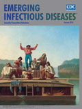
Flatboats, Travelers, Infectious Diseases, and Other River Thoughts [PDF - 288 KB - 2 pages]
| EID | Breedlove B. Flatboats, Travelers, Infectious Diseases, and Other River Thoughts. Emerg Infect Dis. 2016;22(1):162-163. https://doi.org/10.3201/eid2201.ac2201 |
|---|---|
| AMA | Breedlove B. Flatboats, Travelers, Infectious Diseases, and Other River Thoughts. Emerging Infectious Diseases. 2016;22(1):162-163. doi:10.3201/eid2201.ac2201. |
| APA | Breedlove, B. (2016). Flatboats, Travelers, Infectious Diseases, and Other River Thoughts. Emerging Infectious Diseases, 22(1), 162-163. https://doi.org/10.3201/eid2201.ac2201. |


