Rickettsia parkeri Rickettsiosis, Arizona, USA
Kristen L. Herrick, Sandra A. Pena, Hayley Yaglom, Brent J. Layton, Amanda Moors, Amanda D. Loftis, Marah E. Condit, Joseph Singleton, Cecilia Y. Kato, Amy M. Denison, Dianna Ng, James W. Mertins, and Christopher D. Paddock

Author affiliations: Arizona Department of Health Services, Phoenix, Arizona, USA (K.L. Herrick, H.D. Yaglom); Gila County Division of Health and Emergency Management, Globe, Arizona, USA (S.A. Pena); Pinal Mountain Internal Medicine, Globe (B.J. Layton); Moors Wildlife Management Services, Globe (A. Moors); Midwestern University, Glendale, Arizona, USA (A.D. Loftis); Centers for Disease Control and Prevention, Atlanta, Georgia, USA (M.E. Condit, J. Singleton, C.Y. Kato, A.M. Denison, D. Ng, C.D. Paddock); National Veterinary Services Laboratories, Ames, Iowa, USA (J.W. Mertins)
Main Article
Figure 2
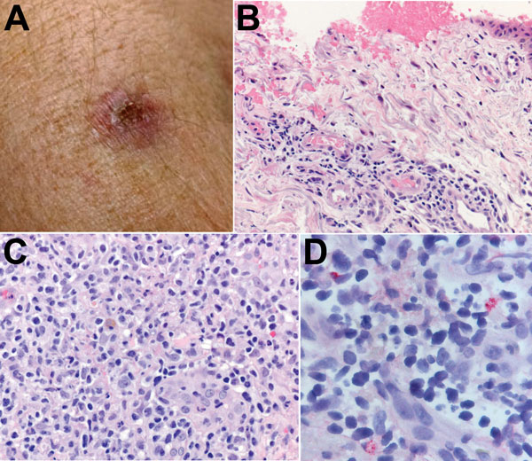
Figure 2. A) Eschar on the right arm of patient 1 at the site of tick bite sustained in Santa Cruz County, Arizona, USA. B) Histological appearance of the eschar biopsy specimen showing ulcerated epidermis with hemorrhage and perivascular lymphohistiocytic inflammatory infiltrates in the superficial dermis. Hematoxylin-eosin staining; original magnification ×50. C) Dense lymphohistiocytic infiltrates around eccrine ducts in the deep dermis of the biopsy specimen. Hematoxylin-eosin staining; original magnification ×100. D) Sparsely distributed intracellular antigens of Rickettsia parkeri (red) within the inflammatory infiltrates, detected by immunohistochemistry. Alkaline phosphatase with naphthol-fast red and hematoxylin counterstaining; original magnification ×158.
Main Article
Page created: April 13, 2016
Page updated: April 13, 2016
Page reviewed: April 13, 2016
The conclusions, findings, and opinions expressed by authors contributing to this journal do not necessarily reflect the official position of the U.S. Department of Health and Human Services, the Public Health Service, the Centers for Disease Control and Prevention, or the authors' affiliated institutions. Use of trade names is for identification only and does not imply endorsement by any of the groups named above.