Volume 22, Number 8—August 2016
CME ACTIVITY - Research
Cutaneous Melioidosis Cluster Caused by Contaminated Wound Irrigation Fluid
Adam J. Merritt, Mariani Peck, Dionne Gayle, Avram Levy, Yi-Horng Ler, Edward Raby, Tristan M. Gibbs, and Timothy J.J. Inglis
Figure 3
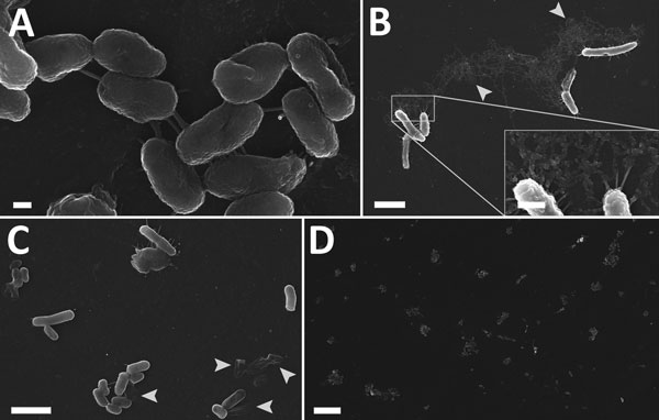
Figure 3. Scanning electron micrographs of internal plastic surface of contaminated irrigation fluid bottle implicated in a 2012–2013 cutaneous melioidosis cluster in the temperate southern region of Western Australia. A–C) Bacilli tethered to each other and to the surface by short peritrichous or polar adhesions (A, C) and occasionally by fibrillary material (B), which appeared to have a globular structure at higher magnification. Decayed cells were common (arrows). D) Clusters of cells were regularly dispersed over the surface. Scale bars indicate 2 μm (A); 200 nm (B and C); 500 nm (B, inset); and 10 μm (D).
Page created: July 08, 2016
Page updated: July 08, 2016
Page reviewed: July 08, 2016
The conclusions, findings, and opinions expressed by authors contributing to this journal do not necessarily reflect the official position of the U.S. Department of Health and Human Services, the Public Health Service, the Centers for Disease Control and Prevention, or the authors' affiliated institutions. Use of trade names is for identification only and does not imply endorsement by any of the groups named above.