Volume 23, Number 11—November 2017
Dispatch
Phylogenetic Analysis of Klebsiella pneumoniae from Hospitalized Children, Pakistan
Figure 1

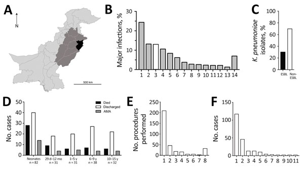
Figure 1. Statistical overview of bacterial isolates from clinical samples collected during May 2010–February 2012 from The Children’s Hospital & The Institute of Child Health, Lahore, Pakistan. A) Map of Pakistan highlighting the main catchment area of Lahore (black, population ≈10 million) and the wider area of Punjab (medium gray, population ≈100 million). B) A total of 5,475 samples collected from children resulted in laboratory-positive cultures; the 5 most frequently occurring bacterial species accounted for ≈70% of total bacterial infections, and Klebsiella pneumoniae (white bar) was the third most dominant (710 isolates). 1, Escherichia coli; 2, coagulase-negative Staphylococcus; 3, K. pneumoniae; 4, Pseudomonas aeruginosa; 5, K. oxytoca; 6, Staphylococcus aureus; 7, Acinetobacter spp.; 8, Enterococcus faecalis; 9, Citrobacter spp.; 10, Streptococcus pyogenes; 11, Burkhoderia cepacia; 12, Enterobacter clocae; 13, Salmonella enterica var. Typhi; 14, others (>100 species). C) The proportion of ESBL-producing K. pneumoniae (214 isolates) among all K. pneumoniae isolates demonstrated high prevalence of antimicrobial resistance. D) A total of 38.3% of ESBL-producing K. pneumoniae infections occurred in neonates (<29 d), an age group that also showed the highest fatality rate (34.1%). Patients who were removed from the hospital against medical advice (AMA) typically were critically ill and were taken home by the family to avoid dying in the hospital. E) The apparent hierarchy shown in panel E closely correlated with interventions given. IV line (97.7%), urinary catheter (27.5%), and ETT (8.4%) were the 3 most commonly administered procedures among sampled patients, although no temporal relationship between procedure and sample collection could be established. 1, IV line; 2, urinary catheter; 3, ETT; 4, PD catheter; 5, surgery; 6, NG tube; 7, CVP; 8, others. F) A total of 54.6% of ESBL-producing K. pneumoniae isolates were from patient blood samples, followed by urine (21.5%), CSF (6%), and ETT (6%). 1, Blood; 2, urine; 3, CSF; 4, ETT; 5, PD catheter; 6, tracheal secretions; 7, pus; 8, CVP tip; 9, ear swab; 10, pleural fluid; 11, wound swab. CSF, cerebrospinal fluid; CVP, central venous catheter tip; ESBL, extended-spectrum β-lactamase; ETT, endotracheal tube; IV, intravenous; NG, nasogastric; PD, peritoneal dialysis catheter. The regional map was derived from the Global Administrative Areas online resource (http://www.gadm.org/).
1These authors contributed equally to this article.
2Current affiliation: CAMS, Aljouf University, Aljouf, Saudi Arabia.