Volume 23, Number 11—November 2017
Dispatch
Mycobacterium lepromatosis Lepromatous Leprosy in US Citizen Who Traveled to Disease-Endemic Areas
Abinash Virk , Bobbi Pritt, Robin Patel, James R. Uhl, Spencer A. Bezalel, Lawrence E. Gibson, Barbara M. Stryjewska, and Margot S. Peters
Figure 2
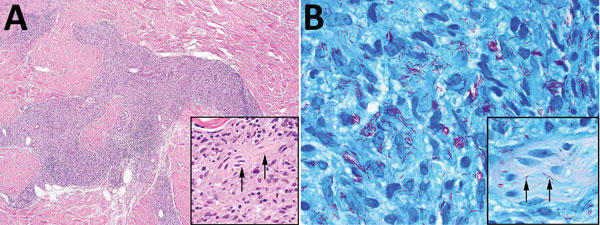
Figure 2. Skin biopsies of 59-year-old white male US citizen showing Mycobacterium lepromatosis infection, 2017. A) Hematoxylin and eosin–stained section of a specimen from the chin showing granulomatous dermal inflammation (original magnification ×100); inset shows nerve involvement (arrows) that is diagnostic for leprosy (original magnification ×400). B) Fite-stained section of a specimen from the chin highlights numerous acid-fast bacilli within histiocytes (original magnification ×1,000); inset shows peripheral nerve involvement (arrows) that is diagnostic for leprosy (original magnification ×1,000).
Page created: October 17, 2017
Page updated: October 17, 2017
Page reviewed: October 17, 2017
The conclusions, findings, and opinions expressed by authors contributing to this journal do not necessarily reflect the official position of the U.S. Department of Health and Human Services, the Public Health Service, the Centers for Disease Control and Prevention, or the authors' affiliated institutions. Use of trade names is for identification only and does not imply endorsement by any of the groups named above.