Volume 23, Number 4—April 2017
Research Letter
Bartonella-Associated Transverse Myelitis
Parham Sendi , Cedric Hirzel, Andreas Bloch, Urs Fischer, Natalie Jeannet, Livia Berlinger, and Heinz Krestel
Figure
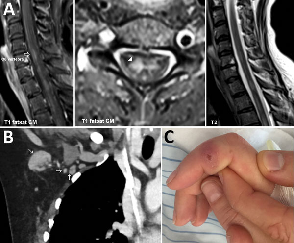
Figure. Images of woman with transverse myelitis and Bartonella henselea infection. A) Magnetic resonance image of the spine showing transverse myelitis (arrowhead). Fat-saturated (fs) T1-weighted image with contrast medium (cm), sagittal plane (left panel) and axial plane (middle panel). T2-weighted image, sagittal plane (right panel). B) Coronal view of computed tomography image of the chest, showing right axillar lymphadenopathy (arrows). C) Right index finger, showing a persistent ulcer from a cat scratch.
Page created: March 17, 2017
Page updated: March 17, 2017
Page reviewed: March 17, 2017
The conclusions, findings, and opinions expressed by authors contributing to this journal do not necessarily reflect the official position of the U.S. Department of Health and Human Services, the Public Health Service, the Centers for Disease Control and Prevention, or the authors' affiliated institutions. Use of trade names is for identification only and does not imply endorsement by any of the groups named above.