Volume 23, Number 6—June 2017
Research Letter
Angiostrongylus cantonensis Meningitis and Myelitis, Texas, USA
Roukaya Al Hammoud , Stacy L. Nayes, James R. Murphy, Gloria P. Heresi, Ian J. Butler, and Norma Pérez
Figure
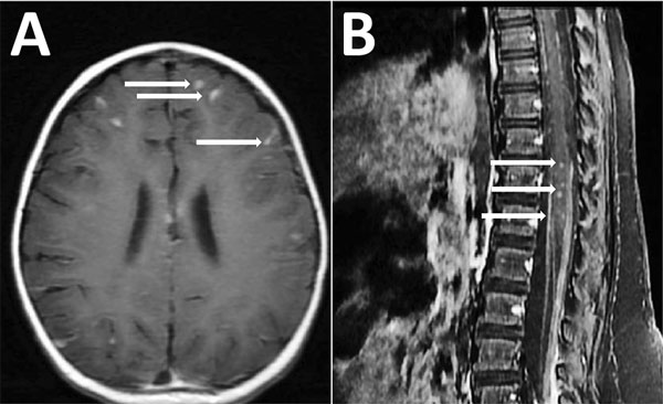
Figure. Magnetic resonance imaging (MRI) of the brain (A) and the spine (B) showing meningitis and myelitis in a 12-month-old girl with Angiostrongylus cantonensis infection, Houston, Texas, USA. A) Axial T1 post contrast sequences showing diffuse leptomeningeal enhancement (arrows). B) Sagittal T1 postcontrast sequences showing intramedullary enhancement in the thoracic and lumbar spinal cord T8–L5 with diffuse leptomeningeal enhancement (arrows).
Page created: May 16, 2017
Page updated: May 16, 2017
Page reviewed: May 16, 2017
The conclusions, findings, and opinions expressed by authors contributing to this journal do not necessarily reflect the official position of the U.S. Department of Health and Human Services, the Public Health Service, the Centers for Disease Control and Prevention, or the authors' affiliated institutions. Use of trade names is for identification only and does not imply endorsement by any of the groups named above.