Volume 23, Number 6—June 2017
Dispatch
Brucella neotomae Infection in Humans, Costa Rica
Figure 1

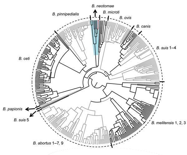
Figure 1. Dendrogram based on multiple locus variable number of tandem repeats–16-loci panel analysis of Brucella spp. (http://mlva.u-psud.fr/brucella/) and clinical isolates from human cerebrospinal fluid samples from 2 patients with brucellosis. The isolates bneohCR1 and bneohCR2 (red branches) showed a pattern consistent with previously reported profiles for Brucella neotomae (blue shading). Black, gray, and tic marks are used to differentiate between adjacent species. Arrows separate small neighboring clusters and indicate the B. neotomae cluster.
1Current affiliation: University of Liverpool, Liverpool, UK
Page created: July 11, 2017
Page updated: July 11, 2017
Page reviewed: July 11, 2017
The conclusions, findings, and opinions expressed by authors contributing to this journal do not necessarily reflect the official position of the U.S. Department of Health and Human Services, the Public Health Service, the Centers for Disease Control and Prevention, or the authors' affiliated institutions. Use of trade names is for identification only and does not imply endorsement by any of the groups named above.