Volume 23, Number 8—August 2017
Research
Molecular Characterization of Corynebacterium diphtheriae Outbreak Isolates, South Africa, March–June 2015
Cite This Article
Citation for Media
Abstract
In 2015, a cluster of respiratory diphtheria cases was reported from KwaZulu-Natal Province in South Africa. By using whole-genome analysis, we characterized 21 Corynebacterium diphtheriae isolates collected from 20 patients and contacts during the outbreak (1 patient was infected with 2 variants of C. diphtheriae). In addition, we included 1 cutaneous isolate, 2 endocarditis isolates, and 2 archived clinical isolates (ca. 1980) for comparison. Two novel lineages were identified, namely, toxigenic sequence type (ST) ST-378 (n = 17) and nontoxigenic ST-395 (n = 3). One archived isolate and the cutaneous isolate were ST-395, suggesting ongoing circulation of this lineage for >30 years. The absence of preexisting molecular sequence data limits drawing conclusions pertaining to the origin of these strains; however, these findings provide baseline genotypic data for future cases and outbreaks. Neither ST has been reported in any other country; this ST appears to be endemic only in South Africa.
Respiratory diphtheria, caused by toxigenic strains of the gram-positive bacillus Corynebacterium diphtheriae, is an upper respiratory tract disease characterized by a sore throat; mild fever; and gray-white pseudomembrane on the tonsils, larynx, or pharynx. Introduction of the diphtheria toxoid vaccine in 1923 and widespread mass immunization in the 1940s and 1950s led to the near elimination of the disease in the industrialized world (1). However, diphtheria remains endemic in many developing countries despite implementation of the World Health Organization Expanded Programme on Immunization in 1974. Epidemic diphtheria resurged in Russia and Eastern Europe in the early 1990s, with >150,000 reported cases occurring predominantly in older children and adults (2).
Molecular epidemiology can be used for investigating diphtheria case clusters in the postvaccine era to improve our understanding of patterns of transmission and spread of epidemic clones. At the time of the Russia epidemic, ribotyping was the established gold standard method of strain typing; however, if deviations from the prescribed protocols occurred, reproducibility could have been compromised (3). Subsequently, multilocus sequence typing (MLST) was developed, which is highly reproducible and provides more accurate information regarding the population structure and evolution (4). More recently, core-genome phylogenetic analyses showed a high level of discrimination and was able to provide insight into C. diphtheriae genomic diversity and identify factors contributing to virulence (5). In addition, the highly discriminatory, clustered, regularly interspaced short palindromic repeats (CRISPR) spoligotyping was used for a more detailed analysis of the Russia epidemic clone (6).
In South Africa, early studies in the 1940s and 1950s reported rates of respiratory diphtheria significantly higher than those in industrialized countries, ranging 20–35 cases/100,000 population and equating to ≈3,000 case notifications annually (7,8). During 1980–2014, a total of 412 diphtheria cases were reported in South Africa through the World Health Organization–UNICEF joint reporting process with most (>80%) notified before 1990 (9). The last laboratory-confirmed respiratory diphtheria case reported in South Africa occurred in a 22-year-old woman in February 2010 in Western Cape Province (10).
During March–June 2015, a cluster of 15 respiratory diphtheria patients with a case-fatality ratio of 27% was reported from KwaZulu-Natal Province in South Africa (11). In 2014, before the outbreak, a KwaZulu-Natal official reported that the province had 96% coverage for the primary series of diphtheria vaccinations and 83% coverage for the 18-month booster (N. McKerrow, KwaZulu-Natal Department of Health, pers. comm., 2015 Jun 8). However, the tetanus-diphtheria booster coverage rates were 54% for 6-year-olds and 20% for 12-year-olds. In response to the outbreak of diphtheria, contact tracing was conducted and postexposure prophylaxis was given to family members and school and clinic contacts (11). Educational leaflets about social mobilization and health promotion activities were distributed in affected communities. The KwaZulu-Natal Department of Health embarked on a catch-up vaccination campaign for schoolgoing children 6–15 years of age in the affected districts. National guidelines for the management and public health response to diphtheria were developed (12), and healthcare practitioners countrywide were notified to be on the alert for possible cases. Laboratories in South Africa were requested to include selective media for isolation of C. diphtheriae when processing throat swabs and to submit all potential C. diphtheriae isolates to the national reference laboratory for confirmation and to establish an isolate repository for molecular surveillance. We conducted a molecular epidemiologic investigation by using whole-genome data to characterize isolates from cases and contacts linked to this KwaZulu-Natal outbreak.
Definitions
We defined a confirmed case as the occurrence of clinical symptoms consistent with respiratory diphtheria (sore throat; low-grade fever; and an adherent membrane on the pharynx, tonsils, larynx, or nose) in a person who was positive for toxin-producing C. diphtheriae and a probable case as the occurrence of mild respiratory symptoms or clinical symptoms of respiratory diphtheria in a C. diphtheriae culture–negative person who was epidemiologically linked to a patient or carrier positive for toxin-producing C. diphtheriae. For the purposes of this investigation, we defined a carrier as a person with a laboratory-confirmed, toxin-producing or non–toxin-producing C. diphtheriae infection with no respiratory symptoms.
Bacterial Strain Collection
During the 2015 KwaZulu-Natal diphtheria outbreak investigation, we received 21 C. diphtheriae isolates swabbed from the throat or nasopharynx (or groin in 1 case). We confirmed identification of cultures by matrix-assisted laser desorption/ionization time-of-flight technology (13) and confirmed the presence of the A and B subunit genes of the C. diphtheriae toxin and phenotypic toxin production by 2 different real-time PCR assays (14,15) and Elek testing (16). Isolates were biotyped with the API Coryne kit (BioMérieux, Lyon, France). We included 2 archived clinical isolates of C. diphtheriae that were isolated in South Africa during the 1980s (although no clinical or demographic data were available for these isolates) and 2 C. diphtheriae isolates from preadolescent children with endocarditis obtained in July and August 2015 (Table 1). Positive controls for PCR and Elek testing were C. diphtheriae vaccine-type strain PW8 (ATCC 13812; American Type Culture Collection, Manassas, VA, USA); toxin-positive C. diphtheriae NCTC 3984 and 10648 (National Collection of Type Cultures, Salisbury, UK); and toxin-negative C. diphtheriae NCTC 10356. Negative controls were C. ulcerans (NCTC 12077), C. bovis (ATCC 7715), and C. striatum (ATCC BAA-1293) (Table 1).
Whole-Genome Sequencing
We extracted DNA from overnight broth cultures with the QIAamp DNA Mini Kit (QIAGEN, Hilden, Germany). We prepared multiplexed paired-end libraries (2 × 300 bp) with the Nextera XT DNA sample preparation kit (Illumina, San Diego, CA, USA) and performed sequencing on an Illumina MiSeq instrument with depth of coverage ranging from 95× to 182×. The raw reads were checked for quality, trimmed, and mapped to the reference genome of C. diphtheriae NCTC 13129 (17) by using CLC Genomics Workbench version 8.5.1 (CLC Bio-QIAGEN, Aarhus, Denmark), which resulted in an 89.7%–93.8% coverage of the reference genome. We performed de novo assembly for all genomes with CLC Genomics and ordered them relative to NCTC 13129 by using the Mauve genome alignment package version 2.3.1 (18). We annotated all genomes by using PROKKA version 1.11 (http://www.vicbioinformatics.com/software.prokka.shtml) and screened the annotated genomes to confirm the presence or absence of the A and B subunits of the C. diphtheriae toxin gene and the toxin repressor gene (dtxR). C. diphtheriae draft genomes for the South Africa isolates have been deposited at DDBJ/European Nucleotide Archive/GenBank (accession nos. MIOA00000000–MIOP00000000, MINX00000000–MINZ00000000, and MIYN00000000–MIYS00000000).
Multilocus Sequence Typing
We retrieved the sequence type (ST) for each isolate from the whole-genome sequence with the Bio-MLST-MLST-Check module (http://search.cpan.org/dist/Bio-MLST-Check/) and applied the eBURST version 3 algorithm (http://eburst.mlst.net/) to generate a population snapshot for C. diphtheriae (19). We defined a clonal complex as a cluster of related STs linked as single-locus variants to another ST in the group. We used all available C. diphtheriae isolates (n = 616) listed in the global MLST database (https://pubmlst.org/cdiphtheriae/) at the time of analysis (accessed June 13, 2017), including 25 isolates from South Africa, to provide context for the South Africa isolates.
Whole-Genome Phylogeny and CRISPR Analysis
We constructed the core-genome alignment by using rapid large-scale prokaryote pan genome analysis (Roary) software (20) to determine the genetic relatedness between the outbreak, outbreak-associated, historical, and endemic isolates. We generated a maximum-likelihood phylogenetic tree by using RaxML version 8 (21). To contextualize the South Africa isolates, we included C. diphtheriae genomes from ATCC and NCTC control strains (Table 1) together with publicly available genomes from Brazil (n = 3) (5), India (n = 2) (22), and Malaysia (n = 2) (23) (selected from GenBank). We identified CRISPR-Cas systems in silico with the online CRISPRFinder program (24) and determined CRISPR-Cas cassettes by using the classification and nomenclature described by Makarova et al. (25).
Ethics
In South Africa, the National Health Act of 2003 (Act No. 61 of 2003) and the Health Professions Act of 1974 (Act No. 56 of 1974) allow access to patient medical records for those working on investigations directed at ensuring the public health. Further, the Human Research Ethics Committee of the University of the Witwatersrand serves the interests of the public in the collection, analysis, and interpretation of communicable disease data. This institution, which has oversight over the National Institute of Communicable Diseases and ensures the application of good clinical and laboratory practices, approved this outbreak investigation (ethics certification no. M160667).
Description of Patients and Contacts with C. diphtheriae
As of June 13, 2015, a total of 15 illnesses were under investigation: 11 were classified as laboratory-confirmed cases and 2 as probable cases (Table 2). One probable case occurred in a patient who was linked to a carrier of toxigenic C. diphtheriae and died; the postmortem throat swab from this patient was culture negative. The second probable case occurred in a patient infected with non–toxin-producing C. diphtheriae who was linked to 3 carriers colonized with toxin-producing C. diphtheriae. The remaining 2 illnesses under investigation could not be classified as confirmed or probable cases; they occurred in culture-negative patients with respiratory diphtheria symptoms who could not be epidemiologically linked to a patient or carrier with toxin-producing C. diphtheriae. Of the 11 patients with laboratory-confirmed cases, 6 patients were not up-to-date with the South African vaccination schedule and 2 had received all scheduled vaccines recommended for their age group. Vaccination status was unknown for 4 patients. With the exception of patient 9, who was white, all other patients and carriers were black.
Among the 292 patients and contacts with throat swab samples taken during the KwaZulu-Natal outbreak investigation, we isolated C. diphtheriae from 19 persons (Table 2). C. diphtheriae isolates from the 11 laboratory-confirmed cases were a mixture of biotypes mitis (n = 8), gravis (n = 1), and intermedius (n = 2). One patient (no. 3) was infected with both toxigenic (biotype mitis) and nontoxigenic (biotype gravis) C. diphtheriae. The 1 culture-positive probable case (no. 14) was defined as such because the patient had respiratory diphtheria symptoms but was culture-positive for nontoxigenic C. diphtheriae. However, this patient had 3 contacts who were carriers (nos. 15, 16, and 17) colonized with toxigenic C. diphtheriae and 1 contact (no. 18) who was a carrier colonized with non–toxin-producing C. diphtheriae. We isolated toxigenic C. diphtheriae from carriers (nos. 7 and 8) who were family members of patients with laboratory-confirmed cases (nos. 5 and 6). One carrier (no. 13) had toxigenic C. diphtheriae and was a contact of another patient with probable diphtheria who died.
C. diphtheriae biotype mitis was predominant among carriers (5/7, 71%). Cutaneous C. diphtheriae biotype gravis was isolated from the groin of a patient (no. 4) who had contact with 2 carriers of non–toxin-producing C. diphtheriae. Isolates from both these carriers were discarded at the source laboratory, and no further characterization of their isolates was possible.
Tox and dtxR Genes
PCR and whole-genome analysis confirmed the results of the phenotypic Elek testing. In addition, all of the South Africa isolates (both toxigenic and nontoxigenic) were found to harbor an intact dtxR gene.
MLSTs and Population Structure
Figure 1
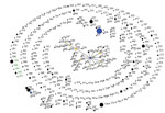
Figure 1. Global population snapshot of Corynebacterium diphtheriae sequence types. eBURST (http://eburst.mlst.net/) was used to display the C. diphtheriae isolates (n = 616) available in the PubMLST database (http://pubmlst.org/cdiphtheriae/) at the time of...
At the time of this analysis, the C. diphtheriae PubMLST database had records from 32 countries dating from 1948 through 2017, with C. diphtheriae isolates from France accounting for 28% of submissions. Overall, the population snapshot revealed a highly diverse population structure for C. diphtheriae globally (Figure 1). The 25 South Africa isolates (21 outbreak-associated and 4 historical) represented 5 novel STs: ST-378 (n = 17), ST-395 (n = 5), ST-390 (n = 1), ST-391 (n = 1), and ST-402 (n = 1). The toxigenic outbreak strain from KwaZulu-Natal was ST-378 and the nontoxigenic strain was unrelated ST-395. The 2 historical isolates from the 1980s (both toxin negative) were ST-395 and unrelated ST-402. The toxin-negative isolates from the endocarditis patients were ST-390 and ST-391 (Table 1), each of which shares 4 of 7 alleles with ST-395.
Core-Genome Phylogeny
Figure 2
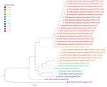
Figure 2. Phylogenetic analysis of Corynebacterium diphtheriae isolates based on sequence type, KwaZulu-Natal Province, South Africa, March–June 2015. Maximum likelihood phylogenetic tree demonstrating core-genome phylogeny among isolates from South Africa (n = 25)...
Consistent with MLST data, we identified 2 distinct lineages among the KwaZulu-Natal outbreak isolates. The 17 toxigenic isolates (from 11 patients and 6 contacts) clustered closely together on the whole-genome phylogenetic tree (Table 2; Figure 2). The second lineage consisted of 5 toxin-negative ST-395 C. diphtheriae isolates: the isolate from the cutaneous diphtheria patient (no. 4), the isolate from the patient with probable diphtheria (no. 14), an isolate from a carrier (no. 18) linked to the patient with probable diphtheria (no. 14), the nontoxigenic isolate from the patient (no. 3) infected with 2 strains of C. diphtheriae, and historical isolate 6853 (Table 1). Comparator genomes from Malaysia (n = 2), Brazil (n = 1), and India (n = 2) and the historical isolate 2337 from South Africa were more closely related to the ST-378 lineage. Two genomes from Brazil (from nontoxigenic isolates) and the UK genome (representative of the Russia outbreak strain) were more closely related to the nontoxigenic ST-395 lineage than to ST-378 (Figure 2; Table 1). The nontoxigenic endocarditis isolates were most closely related to the ST-395 lineage.
CRISPR-Cas Diversity
All of the toxigenic ST-378 isolates harbored a type I-E-a CRISPR-Cas system, with 5 different variants determined by numbers of spacers (Table 2). The 2 carriers (nos. 7 and 8) and 2 patients (nos. 5 and 6) that were from the same family shared an identical CRISPR-Cas type: I-E-a (37 spacers). This specific variant was present in C. diphtheriae from 3 other patients (nos. 3, 10, and 11). The remainder of the ST-378 isolates harbored other type I-E-a variants. Toxigenic C. diphtheriae from carriers from the same family (nos. 15, 16, and 17) harbored identical type I-E-a (31 spacers) CRISPRs.
Toxin-negative ST-395 isolates harbored 2 CRISPR-Cas systems, type I-E-a and type I-E-b, and each of the 4 isolates had their own unique combination of variants. Historical isolate 6853 had CRISPR type I-E-a (12 spacers) and historical isolate 2337 had CRISPR type I-E-b (7 spacers), both of which were unrelated to the KwaZulu-Natal isolates. Similarly, C. diphtheriae from the 2 endocarditis patients had unique CRISPR types I-E-a (47 spacers) and I-E-b (4 spacers).
We describe the molecular epidemiology of C. diphtheriae isolated from a cluster of respiratory diphtheria cases in KwaZulu-Natal, South Africa, during the autumn and winter months of 2015 (11). Suboptimal vaccination coverage rates might have contributed to increased vulnerability of older children and adults to C. diphtheriae infection, leading to this localized cluster of cases. The KwaZulu-Natal outbreak was caused by a single strain, with a novel ST that does not currently belong to any known clonal complex. This toxigenic strain was unrelated to the nontoxigenic strain isolated during this outbreak investigation from the carrier and patients with respiratory and cutaneous diphtheria. The strain was also not related to the historical and non–outbreak-associated isolates from South Africa or to any other documented C. diphtheriae ST from elsewhere in the world.
Biotype does not appear associated with disease severity and is regarded as having limited utility in epidemiologic investigations because of poor discrimination and lack of correlation with genotype (4,26). Nevertheless, a shift in the predominant circulating biotype of C. diphtheriae was demonstrated during a Russia outbreak (27,28). However, of the 2 studies on the Russia outbreak, neither had genome data available for comparison. In our investigation, no relationship between biotype and genotype was evident. In addition, no epidemiologic correlation with biotype was apparent, as demonstrated by 4 members of the same family being infected with identical C. diphtheriae genotypes but having different biotypes (mitis and gravis).
MLST was consistent with core-genome phylogeny: isolates of the same ST clustered together. In addition to the MLST and core-genome phylogeny, analysis of the CRISPR systems provided additional resolution between the 2 KwaZulu-Natal lineages and allowed a better understanding of the transmission dynamics. Finding C. diphtheriae with identical CRISPR variants among family members confirmed circulation and transmission of the same strain. Sequence errors might have resulted in misclassification of some variants that were not confirmed by another method or repeat sequencing; nevertheless, a good depth of sequencing coverage together with other parameters validated the quality of the reads and accuracy of base calling.
C. diphtheriae has been shown to exhibit a high degree of genome plasticity (29), which was reflected by the highly diverse global population (consisting of mostly unrelated genotypes) seen with the STs from PubMLST. Individual population snapshots by country reiterate this heterogeneity, and many STs are unique to their respective countries (data not shown). One dominant clone ST-42 and its associated single and double-locus variants, which were predominantly isolated in France, accounted for 7% of C. diphtheriae isolates in PubMLST; however, the global database is overrepresented by submissions from France and, thus, might not accurately reflect the true population. The MLST database includes a mixture of unrelated STs from countries of North and West Africa, and Angola (the geographically closest neighbor to South Africa for which data were available) has reported a single ST-316 isolate. None of the isolates from Africa share the same or related STs as those identified in the outbreak we report. MLST data for C. diphtheriae isolates collected over >20 years (1992–2015) in Algeria showed most isolates were ST-116 (unrelated to any other ST in the PubMLST database), the predominant ST circulating during the 1992–1999 epidemic in that country (30). In later years, the population structure of C. diphtheriae in Algeria was more heterogeneous.
The South Africa outbreak-associated isolates represented 2 novel STs not previously reported or related to any other PubMLST-listed ST in the global database. The lack of data for C. diphtheriae from other countries in Africa makes it difficult to speculate whether these strains were imported from close neighboring countries or farther away, or whether they are endemic to South Africa. Subsequent to the 2015 cluster of cases in South Africa, we also reported 2 additional cases in May 2016 in the same region, and both were ST-378.
During the KwaZulu-Natal outbreak, co-circulation of the toxigenic and nontoxigenic genotypically unrelated strains were noted during laboratory investigations. Both genotypes (ST-378 and ST-395) were detected in patients and carriers. In 1 case, 2 C. diphtheriae isolates (1 toxigenic, 1 nontoxigenic) were detected from the same patient. Because CRISPR analysis indicated that the nontoxigenic isolate had a distinctive combination of CRISPR variants not seen in any of the other isolates, the possibility of contamination was excluded. We expect that disease in this patient was caused by the toxigenic strain and not the nontoxigenic strain. In 1 probable case, a person with symptoms clinically suggestive of diphtheria was infected with the nontoxigenic ST-395 strain but was epidemiologically linked to 3 asymptomatic persons colonized with the ST-378 toxigenic strain. In 1978, a similar phenomenon occurred with a 25-year-old woman in Toronto; 3 different C. diphtheriae variants exhibiting 2 different phage types and a mixture of toxigenic and nontoxigenic isolates were isolated from her throat (31). In our study, sampling and testing methods might not have been sensitive enough to detect all C. diphtheriae variants present. Laboratory workers should be aware of the possibility of mixed C. diphtheriae populations in a single person. Although mixtures might be evident through differences in colony and microscopic morphologies, molecular characterization might be more sensitive in detecting such variants.
Cases of nontoxigenic and cutaneous C. diphtheriae might not be clinically as important as toxigenic and respiratory diphtheria and are not notifiable. However, skin lesions can serve as a reservoir of strains that are toxigenic or strains that could potentially become toxigenic if the bacteria possess functional toxin repressor genes and become infected with a tox gene–bearing lysogenic corynephage (32,33). In addition, countries with high vaccination coverage have reported the emergence of invasive disease caused by nontoxigenic C. diphtheriae, particularly in high-risk groups such as persons who are homeless, persons who use intravenous drugs or alcohol, persons with diabetes mellitus, and persons with dental caries (34–36). The South Africa isolates from the 2015 KwaZulu-Natal outbreak were not derived from a single lineage; 2 distinct genotypes were identified within the community. The nontoxigenic lineage has been in circulation for >30 years, verified by sequence typing and close phylogenomic clustering of the archived 1980s isolate (6853) with the ST-395 KwaZulu-Natal outbreak-associated isolates. Because no respiratory or cutaneous diphtheria cases have been reported for several years in South Africa, we have no baseline genotypic data regarding the underlying population diversity, and hence, we are unable to track transmission patterns or changes in genotypes over time. The nontoxigenic South Africa isolates in this study all harbored dtxR genes, indicating the potential for toxin production if lysogenized by a bacteriophage.
We identified 2 novel strains of C. diphtheriae in a province of South Africa during an outbreak investigation occurring >30 years after diphtheria ceased to be a public health concern. The absence of preexisting molecular sequence data limits conclusions pertaining to the origin of these strains; however, these findings provide baseline genotypic data for future cases and outbreaks as well as information on transmission dynamics for the 2015 outbreak. A better understanding of the molecular epidemiology of this pathogen might assist in directing and strengthening public health interventions. Active and passive surveillance for diphtheria and C. diphtheriae carriage is required locally and on the subcontinent, particularly in the context of suboptimal vaccination coverage, especially in older children. We are exploring the idea of a serosurvey in South Africa among different age groups, which might provide insight into the epidemiology of diphtheria in this country.
Dr. du Plessis is a medical laboratory scientist for the Centre for Respiratory Diseases and Meningitis at the National Institute for Communicable Diseases, South Africa, and holds a joint appointment as lecturer at the University of the Witwatersrand, South Africa. Her research interests include the molecular epidemiology of respiratory pathogens and exploring new molecular diagnostic platforms.
Acknowledgments
We would like to acknowledge the tireless efforts of our public health colleagues in KwaZulu-Natal, who were instrumental in confirming cases and coordinating the public health response during the outbreak: Premi Govender and the KwaZulu-Natal Department of Health (DoH) staff; Vusi Mbuthu, Nokuthula Ndlovu, Ayodeji Olowolagba and team, and the Ethekwini Metro DoH staff; Nelly Gumede, Sharon Fynn, Zohra Banoo and team, and the Ethekwini DoH staff; and Siphumelele Mlambo and the Ugu District DoH staff. In addition, we thank all clinicians who were involved in treating patients.
Laboratory testing and molecular characterization was funded by the National Institute for Communicable Diseases.
References
- Efstratiou A, George RC. Microbiology and epidemiology of diphtheria. Rev Med Microbiol. 1996;7:31–42. DOIGoogle Scholar
- Vitek CR, Wharton M. Diphtheria in the former Soviet Union: reemergence of a pandemic disease. Emerg Infect Dis. 1998;4:539–50. DOIPubMedGoogle Scholar
- Dallman T, Neal S, Green J, Efstratiou A. Development of an online database for diphtheria molecular epidemiology under the remit of the DIPNET project. Euro Surveill. 2008;13:18865.PubMedGoogle Scholar
- Bolt F, Cassiday P, Tondella ML, Dezoysa A, Efstratiou A, Sing A, et al. Multilocus sequence typing identifies evidence for recombination and two distinct lineages of Corynebacterium diphtheriae. J Clin Microbiol. 2010;48:4177–85. DOIPubMedGoogle Scholar
- Trost E, Blom J, Soares SC, Huang I-H, Al-Dilaimi A, Schröder J, et al. Pangenomic study of Corynebacterium diphtheriae that provides insights into the genomic diversity of pathogenic isolates from cases of classical diphtheria, endocarditis, and pneumonia. J Bacteriol. 2012;194:3199–215. DOIPubMedGoogle Scholar
- Mokrousov I, Narvskaya O, Limeschenko E, Vyazovaya A. Efficient discrimination within a Corynebacterium diphtheriae epidemic clonal group by a novel macroarray-based method. J Clin Microbiol. 2005;43:1662–8. DOIPubMedGoogle Scholar
- Bokkenheuser V, Heymann CS. Diphtheria in South Africa. S Afr Med J. 1954;28:685–9.PubMedGoogle Scholar
- Bokkenheuser V. Geographical and racial distribution of diphtheria in South Africa. S Afr Med J. 1961;35:711–5.PubMedGoogle Scholar
- World Health Organization. Diphtheria reported cases [cited 2016 Aug 22]. http://apps.who.int/immunization_monitoring/globalsummary/timeseries/tsincidencediphtheria.html
- National Institute for Communicable Diseases. Diphtheria. National Institute for Communicable Diseases Communique. 2010;9(2):4–5 [cited 2016 Jul 25]. http://www.nicd.ac.za/assets/files/NICDCommFeb10Vol09_02.pdf
- Mahomed S, Archary M, Mutevedzi P, Mahabeer Y, Govender P, Ntshoe G, et al. An isolated outbreak of diphtheria in South Africa, 2015. Epidemiol Infect. 2017;1–9.DOIPubMedGoogle Scholar
- Cohen C, de Gouveia L, du Plessis M, McCarthy K, Mlisana K, Moodley P, et al. National Institute for Communicable Diseases. Diphtheria: NICD recommendations for diagnosis, management and public health response. Version 2.3. 2016 Mar 22 [cited 25 July 2016]. http://www.nicd.ac.za/assets/files/Guidelines_diphtheria_20160322_v2_3.pdf
- Konrad R, Berger A, Huber I, Boschert V, Hörmansdorfer S, Busch U, et al. Matrix-assisted laser desorption/ionisation time-of-flight (MALDI-TOF) mass spectrometry as a tool for rapid diagnosis of potentially toxigenic Corynebacterium species in the laboratory management of diphtheria-associated bacteria. Euro Surveill. 2010;15:19699.PubMedGoogle Scholar
- Mancini F, Monaco M, Pataracchia M, von Hunolstein C, Pantosti A, Ciervo A. Identification and molecular discrimination of toxigenic and nontoxigenic diphtheria Corynebacterium strains by combined real-time polymerase chain reaction assays. Diagn Microbiol Infect Dis. 2012;73:111–20. DOIPubMedGoogle Scholar
- Mothershed EA, Cassiday PK, Pierson K, Mayer LW, Popovic T. Development of a real-time fluorescence PCR assay for rapid detection of the diphtheria toxin gene. J Clin Microbiol. 2002;40:4713–9. DOIPubMedGoogle Scholar
- Efstratiou A, Maple PAC. WHO manual for the laboratory diagnosis of diphtheria. Document ICP-EPI 038 (C). Geneva: World Health Organization; 1994.
- Cerdeño-Tárraga AM, Efstratiou A, Dover LG, Holden MT, Pallen M, Bentley SD, et al. The complete genome sequence and analysis of Corynebacterium diphtheriae NCTC13129. Nucleic Acids Res. 2003;31:6516–23. DOIPubMedGoogle Scholar
- Rissman AI, Mau B, Biehl BS, Darling AE, Glasner JD, Perna NT. Reordering contigs of draft genomes using the Mauve aligner. Bioinformatics. 2009;25:2071–3. DOIPubMedGoogle Scholar
- Feil EJ, Li BC, Aanensen DM, Hanage WP, Spratt BG. eBURST: inferring patterns of evolutionary descent among clusters of related bacterial genotypes from multilocus sequence typing data. J Bacteriol. 2004;186:1518–30. DOIPubMedGoogle Scholar
- Page AJ, Cummins CA, Hunt M, Wong VK, Reuter S, Holden MT, et al. Roary: rapid large-scale prokaryote pan genome analysis. Bioinformatics. 2015;31:3691–3. DOIPubMedGoogle Scholar
- Stamatakis A. RAxML version 8: a tool for phylogenetic analysis and post-analysis of large phylogenies. Bioinformatics. 2014;30:1312–3. DOIPubMedGoogle Scholar
- Veeraraghavan B, Anandan S, Rajamani Sekar SK, Gopi R, Devanga Ragupathi NK, Ramesh S, et al. First report on the draft genome sequences of Cornebacterium diphtheriae isolates from India. Genome Announc. 2016;4:e01316-16. DOIPubMedGoogle Scholar
- Ahmad N, Hii FSY, Mohd Khalid MKN, Abd Wahab MA, Hashim R, Tang SN, et al. First draft genome sequences of Malaysian clinical isolates of Cornebacterium diphtheriae. Genome Announc. 2017;5:e016701-16. DOIGoogle Scholar
- Grissa I, Vergnaud G, Pourcel C. CRISPRFinder: a web tool to identify clustered regularly interspaced short palindromic repeats. Nucleic Acids Res. 2007;35(Web Server):W52-7. DOIPubMedGoogle Scholar
- Makarova KS, Haft DH, Barrangou R, Brouns SJ, Charpentier E, Horvath P, et al. Evolution and classification of the CRISPR-Cas systems. Nat Rev Microbiol. 2011;9:467–77. DOIPubMedGoogle Scholar
- Sangal V, Burkovski A, Hunt AC, Edwards B, Blom J, Hoskisson PA. A lack of genetic basis for biovar differentiation in clinically important Corynebacterium diphtheriae from whole genome sequencing. Infect Genet Evol. 2014;21:54–7. DOIPubMedGoogle Scholar
- Kolodkina V, Titov L, Sharapa T, Grimont F, Grimont PA, Efstratiou A. Molecular epidemiology of C. diphtheriae strains during different phases of the diphtheria epidemic in Belarus. BMC Infect Dis. 2006;6:129. DOIPubMedGoogle Scholar
- Markina SS, Maksimova NM, Vitek CR, Bogatyreva EY, Monisov AA. Diphtheria in the Russian Federation in the 1990s. J Infect Dis. 2000;181(Suppl 1):S27–34. DOIPubMedGoogle Scholar
- Iwaki M, Komiya T, Yamamoto A, Ishiwa A, Nagata N, Arakawa Y, et al. Genome organization and pathogenicity of Corynebacterium diphtheriae C7(-) and PW8 strains. Infect Immun. 2010;78:3791–800. DOIPubMedGoogle Scholar
- Benamrouche N, Hasnaoui S, Badell E, Guettou B, Lazri M, Guiso N, et al. Microbiological and molecular characterization of Corynebacterium diphtheriae isolated in Algeria between 1992 and 2015. Clin Microbiol Infect. 2016;22:1005.e1–7. DOIPubMedGoogle Scholar
- Chang DN, Laughren GS, Chalvardjian NE. Three variants of Corynebacterium diphtheriae subsp. mitis (Belfanti) isolated from a throat specimen. J Clin Microbiol. 1978;8:767–8.PubMedGoogle Scholar
- Belsey MA, LeBlanc DR. Skin infections and the epidemiology of diphtheria: acquisition and persistence of C diphtheriae infections. Am J Epidemiol. 1975;102:179–84. DOIPubMedGoogle Scholar
- De Zoysa A, Efstratiou A, Hawkey PM. Molecular characterization of diphtheria toxin repressor (dtxR) genes present in nontoxigenic Corynebacterium diphtheriae strains isolated in the United Kingdom. J Clin Microbiol. 2005;43:223–8. DOIPubMedGoogle Scholar
- Farfour E, Badell E, Zasada A, Hotzel H, Tomaso H, Guillot S, et al. Characterization and comparison of invasive Corynebacterium diphtheriae isolates from France and Poland. J Clin Microbiol. 2012;50:173–5. DOIPubMedGoogle Scholar
- Romney MG, Roscoe DL, Bernard K, Lai S, Efstratiou A, Clarke AM. Emergence of an invasive clone of nontoxigenic Corynebacterium diphtheriae in the urban poor population of Vancouver, Canada. J Clin Microbiol. 2006;44:1625–9. DOIPubMedGoogle Scholar
- Zasada AA. Nontoxigenic highly pathogenic clone of Corynebacterium diphtheriae, Poland, 2004-2012. Emerg Infect Dis. 2013;19:1870–2. DOIPubMedGoogle Scholar
Figures
Tables
Cite This ArticleTable of Contents – Volume 23, Number 8—August 2017
| EID Search Options |
|---|
|
|
|
|
|
|
Please use the form below to submit correspondence to the authors or contact them at the following address:
Mignon du Plessis, Centre for Respiratory Diseases and Meningitis, 1 Modderfontein Rd, Sandringham, Johannesburg 2031 South Africa
Top