Volume 23, Number 8—August 2017
Research
Molecular Characterization of Corynebacterium diphtheriae Outbreak Isolates, South Africa, March–June 2015
Mignon du Plessis , Nicole Wolter, Mushal Allam, Linda de Gouveia, Fahima Moosa, Genevie Ntshoe, Lucille Blumberg, Cheryl Cohen, Marshagne Smith, Portia Mutevedzi, Juno Thomas, Valentino Horne, Prashini Moodley, Moherndran Archary, Yesholata Mahabeer, Saajida Mahomed, Warren Kuhn, Koleka Mlisana, Kerrigan McCarthy, and Anne von Gottberg
Figure 1
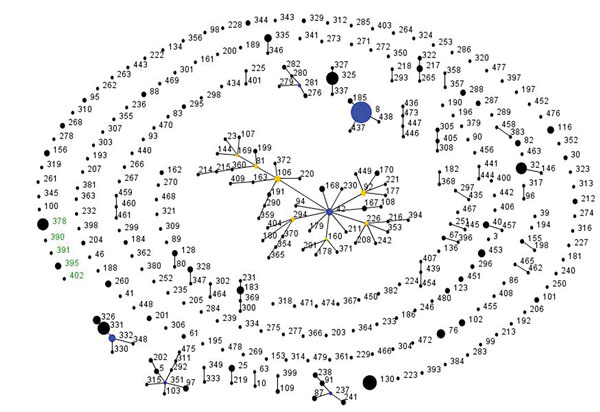
Figure 1. Global population snapshot of Corynebacterium diphtheriae sequence types. eBURST (http://eburst.mlst.net/) was used to display the C. diphtheriae isolates (n = 616) available in the PubMLST database (http://pubmlst.org/cdiphtheriae/) at the time of analysis (accessed June 13, 2017). Isolates from South Africa (n = 25) are green. The size of each circle is proportional to the number of isolates, and related sequence types are connected by lines. Blue and yellow circles indicate founding and subfounding genotypes, respectively.
Page created: July 17, 2017
Page updated: July 17, 2017
Page reviewed: July 17, 2017
The conclusions, findings, and opinions expressed by authors contributing to this journal do not necessarily reflect the official position of the U.S. Department of Health and Human Services, the Public Health Service, the Centers for Disease Control and Prevention, or the authors' affiliated institutions. Use of trade names is for identification only and does not imply endorsement by any of the groups named above.