Volume 23, Number 9—September 2017
Dispatch
Group A Rotavirus Associated with Encephalitis in Red Fox
Figure 1
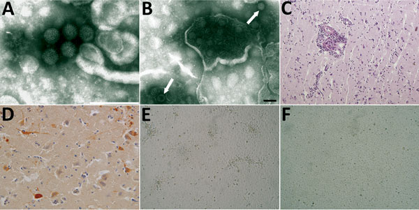
Figure 1. Images of brain of fox with group A rotavirus infection and brains of suckling and weanling mice inoculated with fox brain homogenates. A, B) Negative-staining electron microscopy. Presence of virions morphologically related to family Reoviridae from fox (A) and mouse (B) brain (arrows). Scale bar in panel A indicates 200 nm; in panel B, 100 nm. C, D) Histologic and immunohistochemical appearance of the cerebral cortex of the fox. C) Perivascular cuffing of inflammatory cells in the brain stained by hematoxylin and eosin (original magnification ×10). D) Viral antigen in the cytoplasm of neurons (immunohistochemistry, original magnification ×20). E) Foci with rounding cells of the confluent monolayers of Marc-145 cells infected with the brain homogenate from mouse at 2 days after inoculation (original magnification ×40). F) Mock cells (original magnification ×40).
1Deceased.