Candida auris in Healthcare Facilities, New York, USA, 2013–2017
Eleanor Adams, Monica Quinn, Sharon Tsay, Eugenie Poirot, Sudha Chaturvedi, Karen Southwick, Jane Greenko, Rafael Fernandez, Alex Kallen, Snigdha Vallabhaneni, Valerie Haley, Brad Hutton, Debra Blog, Emily Lutterloh

, Howard Zucker, and Candida auris Investigation Workgroup
1
Author affiliations: New York State Department of Health, New Rochelle, New York, USA (E. Adams, K. Southwick); New York State Department of Health, Albany, New York, USA (M. Quinn, S. Chaturvedi, V. Haley, B. Hutton, D. Blog, E. Lutterloh, H. Zucker); Centers for Disease Control and Prevention, Atlanta, Georgia, USA (S. Tsay, E. Poirot, A. Kallen, S. Vallabhaneni); New York City Department of Health and Mental Hygiene, New York, New York, USA (E. Poirot); New York State Department of Health, Central Islip, New York, USA (J. Greenko); New York State Department of Health, New York (R. Fernandez); State University at Albany School of Public Health, Albany, New York, USA (V. Haley, D. Blog, E. Lutterloh)
Main Article
Figure 3
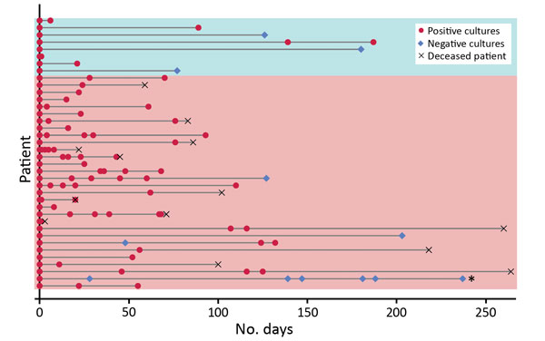
Figure 3. Long-term Candida auris colonization of clinical and screening case-patients, New York, USA, 2013–2017. Each patient for whom follow-up cultures were performed is represented by a horizontal line. The bottom 30 lines (pink shading) indicate clinical case-patients; the top 8 (blue shading) indicate screening case-patients. Follow-up cultures were collected from a variety of sites, typically axilla and groin and often nares, rectum, urine, and wounds. Persons were considered free of colonization with C. auris and eligible for removal of contact precautions when 2 sets of surveillance cultures at multiple sites, taken at least 1 week apart, were negative; only 1 person indicated on the figure (second from bottom) met this criterion.
Main Article
Page created: September 12, 2018
Page updated: September 12, 2018
Page reviewed: September 12, 2018
The conclusions, findings, and opinions expressed by authors contributing to this journal do not necessarily reflect the official position of the U.S. Department of Health and Human Services, the Public Health Service, the Centers for Disease Control and Prevention, or the authors' affiliated institutions. Use of trade names is for identification only and does not imply endorsement by any of the groups named above.