Volume 24, Number 11—November 2018
Research Letter
Congenital Zika Virus Infection with Normal Neurodevelopmental Outcome, Brazil
Alessandra Lemos de Carvalho , Carlos Brites, Tânia Barreto Taguchi, Suely Fernandes Pinho, Gúbio Campos, and Rita Lucena
Figure
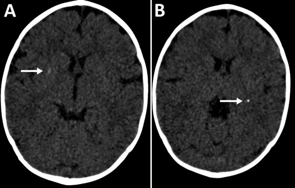
Figure. Cerebral computed tomography images of infant with probable congenital Zika virus infection at 7 months of age, Brazil. A) Mild calcification at the right lenticular nucleus (arrow); B) calcifications at the posterior arm of the left internal capsule (arrow).
Page created: October 17, 2018
Page updated: October 17, 2018
Page reviewed: October 17, 2018
The conclusions, findings, and opinions expressed by authors contributing to this journal do not necessarily reflect the official position of the U.S. Department of Health and Human Services, the Public Health Service, the Centers for Disease Control and Prevention, or the authors' affiliated institutions. Use of trade names is for identification only and does not imply endorsement by any of the groups named above.